
Filamentous Fungi
Overview
Source: Laboratories of Dr. Ian Pepper and Dr. Charles Gerba - The University of Arizona
Demonstrating Author: Bradley Schmitz
Fungi are heterotrophic eukaryotic organisms, and with the exception of yeasts, are aerobic. They are abundant in surface soils and are important for their role in nutrient cycling and the decomposition of organic matter and organic contaminants. White rot fungi (phanerochaete chryosporium) for example, (Figure 1) are known to degrade aromatics.
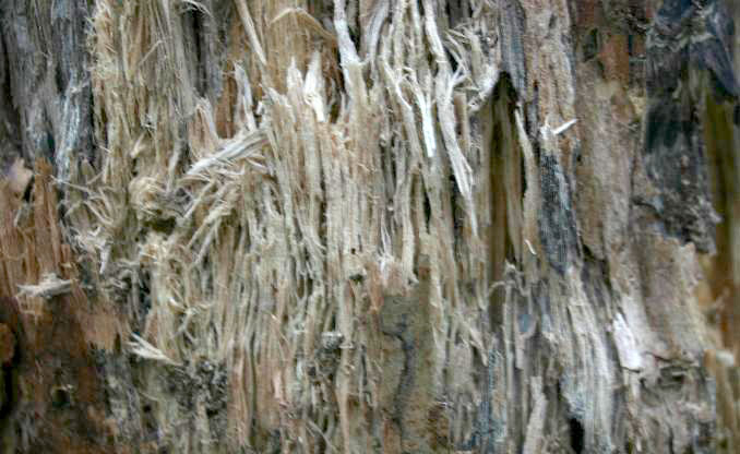
Figure 1. White rot on birch.
Principles
Soils generally contain millions of fungi per gram, so the soil is typically diluted using a dilution series. A dilution series is made by suspending a given amount of soil in a dispersing solution, such as deionized water. The aliquots of the suspensions are then transferred to fresh solution, until the suspension is diluted sufficiently to allow individual discrete fungal colonies to grow on the agar plates. (Figure 2)
After inoculation on several replicate agar plates, the plates are incubated at 25 °C. After the macroscopic fungal colonies are formed, they are counted, as shown in Figure 3. Because the assumption is that one fungal colony is derived from one organism, the term Colony Forming Units (CFUs) is used in the final analysis, with the results expressed in terms of CFUs per gram of oven dry soil.
Normal culturable fungal counts from fertile soil are in the range of 106-106 fungal “propagules” (spores, hyphae, or hyphal fragments) per gram of dry soil. Culturable plate counts have been in use for enumerating organisms since the nineteenth century. They continue to be used today, as they are inexpensive to perform, require little labor, are quick, and are fairly reproducible. However, they do suffer from a number of errors, which must be considered when evaluating the results. The most significant of these errors is the fact that many organisms will not culture on media plates.

Figure 2. Dilution and plating technique. Here, the diluted soil suspension is incorporated directly in the agar medium rather than being surface applied as in the case of spread plating. From Environmental & Pollution Science, 2nd Ed., Academic Press, San Diego, CA
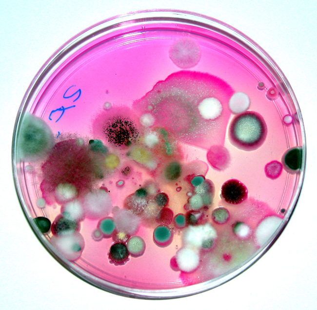
Figure 3. Soil fungi isolated from a surface soil grown in a Petri dish containing Rose Bengal Agar. Photo courtesy K.L. Josephson. From Environmental & Pollution Science, 2nd Ed., Academic Press, San Diego, CA.
Procedure
1. Soil Sample Preparation
- First, determine the initial moisture content of the soil by overnight drying of a known amount of the moist soil, and reweighing the dried soil. The equation to determine the initial moisture content of the soil is:

(Equation 1)
where:
MC = moisture content
W = net weight
D = dry weight - Calculate the amount of water that must be added to 25 g of soil to increase the soil moisture content to 10% on a dry weight basis.
- Next, add that amount of water to 25 g of the soil.
- Cover the container with plastic wrap and puncture the film several times with a probe to allow aeration during incubation. Secure the film with a rubber band.
- Weigh the soil and wrap, then incubate the sample at room temperature for one week.
2. Fungus Inoculation and Incubation
- After the incubation is complete, weigh the soil sample with the wrap and rubber band. Calculate the weight loss due to moisture loss.
- Calculate the new soil moisture content.
- Next, prepare a 1/10-dilution series of the soils as shown in Figure 2. This will result in dilutions of 10-1 (bottle A), 10-2 (tube B), 10-3 (tube C), 10-4 (tube D), and 10-5 (tube E) g soil per mL suspensions.
- Prepare sterile Rose Bengal-streptomycin agar plates. Both the Rose Bengal and streptomycin inhibit bacterial growth. For very fertile soils where soil microbial populations are high, the chosen dilutions should be higher, i.e., 10-3, 10-4, and 10-5, g soil per plate.
- To each of the 10-3 plates, add 0.1 mL from dilution tube 10-2, and spread over the agar surface using an ethanol-flame sterilized glass spreader. Because a quantity of 0.1 mL is plated, this makes the effective dilution 10-3. Repeat these steps for all dilutions.
- Incubate plates at room temperature for one week.
3. Colony Counting and Examination by Microscopy
- Make colony counts at one and only one dilution of each soil. The plates that are counted should have discreet countable colonies. Overgrown plates should not be counted. Likewise, plates with less than 10 colonies should not be counted. Note and describe the cultural characteristics of three different colonies.
- Prepare pressure tape (transparent tape) mounts on slides for detailed microscope study using the following procedure:
- Deposit a drop of lactophenol mounting fluid at the center of a clean glass slide
- Cut a strip of clear cellophane tape about 3 cm long from the stock roll. To avoid contaminating the adhesive surface, use forceps when handling the tape. A dissecting needle will aid in freeing the tape from the forceps.
- The adhesive side of the tape is applied to the surface of a sporulating fungus colony. Take care to avoid excessive pressure on the tape or too dense a mass of hyphae and spores will be collected.
- Remove the tape from contact with the fungus colony and apply it, adhesive side down, to the drop of mounting fluid on the glass slide. Rub the tape gently with a smooth, flat instrument to express air bubbles.
- Examine the tape mount microscopically under the oil immersion objective with oil.
- Identify two different fungal genera using the supplied fungal identification key (Figure 4).

Figure 4. Fungal identification key.
Results
Colony Counts
The number of fungal colonies per gram of soil is equal to the number of colonies counted on the plate multiplied by the reciprocal of the dilution plated. For example, if 46 colonies are counted at a dilution of 10-5, then the CFU per gram of soil is 46 x 105 or 4.6 x 10⁶.
Identification of Three Different Fungal Genera
Fungi can be identified microscopically by examination of the fruiting bodies and spores. A fungi identification key can assist this process. (Figure 4) Common fungi types observed include Penicillium and Aspergillus.
Application and Summary
Dilution and plating of soil fungi can be used as an indication of the health of a soil. Normally a “healthy” fertile soil will have 105 to 106 fungi per gram of soil. It can also be utilized to isolate pure cultures of specific fungi, subsequently evaluated for specific properties, such as the ability to degrade organic compounds. These can be detrimental as in the case of white rot fungi, or beneficial when toxic organics are degraded through biodegradation. Other uses of pure cultures of fungi include the isolation of fungi for antibiotics. For example, the first antibiotic ever was penicillin, produced by the soil-borne fungus Penicillium. This was first discovered by Sir. Alexander Fleming in 1929.
References
- Pepper, I.L., Gerba, C.P., Brusseau, M. Environmental & Pollution Science, 2nd Ed. Academic Press, San Diego, CA. (2006).
- Pepper, I.L., Gerba, C.P. Environmental Microbiology, A Laboratory Manual, 2nd Ed. Academic Press, Boston, MA. (2005).
Tags
Skip to...
Videos from this collection:

Now Playing
Filamentous Fungi
Environmental Microbiology
57.3K Views

Determination of Moisture Content in Soil
Environmental Microbiology
359.3K Views

Aseptic Technique in Environmental Science
Environmental Microbiology
126.4K Views

Gram Staining of Bacteria from Environmental Sources
Environmental Microbiology
100.2K Views

Visualizing Soil Microorganisms via the Contact Slide Assay and Microscopy
Environmental Microbiology
42.2K Views

Community DNA Extraction from Bacterial Colonies
Environmental Microbiology
28.8K Views

Detecting Environmental Microorganisms with the Polymerase Chain Reaction and Gel Electrophoresis
Environmental Microbiology
44.5K Views

RNA Analysis of Environmental Samples Using RT-PCR
Environmental Microbiology
40.4K Views

Quantifying Environmental Microorganisms and Viruses Using qPCR
Environmental Microbiology
47.8K Views

Water Quality Analysis via Indicator Organisms
Environmental Microbiology
29.5K Views

Isolation of Fecal Bacteria from Water Samples by Filtration
Environmental Microbiology
39.3K Views

Detection of Bacteriophages in Environmental Samples
Environmental Microbiology
40.7K Views

Culturing and Enumerating Bacteria from Soil Samples
Environmental Microbiology
184.3K Views

Bacterial Growth Curve Analysis and its Environmental Applications
Environmental Microbiology
295.9K Views

Algae Enumeration via Culturable Methodology
Environmental Microbiology
13.8K Views
Copyright © 2025 MyJoVE Corporation. All rights reserved