Method Article
각막 상피 창상 치유 기간 동안 열 충격 단백질 27의 기능의 RNA 간섭 기반 조사
요약
Herein, we present a protocol to use heat shock protein 27 (HSP27)-specific small interfering RNA to assess the function of HSP27 during corneal epithelial wound healing. RNA interference is the best method for effectively knocking-down gene expression to investigate protein function in various cell types.
초록
Small interfering RNA (siRNA) is among the most widely used RNA interference methods for the short-term silencing of protein-coding genes. siRNA is a synthetic RNA duplex created to specifically target a mRNA transcript to induce its degradation and it has been used to identify novel pathways in various cellular processes. Few reports exist regarding the role of phosphorylated heat shock protein 27 (HSP27) in corneal epithelial wound healing. Herein, cultured human corneal epithelial cells were divided into a scrambled control-siRNA transfected group and a HSP27-specific siRNA-transfected group. Scratch-induced directional wounding assays, and western blotting, and flow cytometry were then performed. We conclude that HSP27 has roles in corneal epithelial wound healing that may involve epithelial cell apoptosis and migration. Here, step-by-step descriptions of sample preparation and the study protocol are provided.
서문
그들은 동시에 윤부 각막 상피 기저 층에서 세포로 대체하는 동안 각막 상피 세포 (CECs)이 지속적으로, 눈물 막으로 흘려된다. (1) 다양한 외부 스트레스가 CECs의 세포 사멸 및 박리를 유도 할 수 있습니다. 2 열 충격 단백질들 (HSPs) 고도로 보존되어 있으며, 분자의 크기에 따라 두 개 패밀리로 분할 될 수있다. (3) 최대 HSP 계열은 HSP90, HSP70, 및 HSP60 및 작은 가족 HSP27 포함을 포함한다. 4 HSP27의 인산화는 세포 생존에 중요한 역할을하는 것으로 알려져 때문에 굴지의 리모델링이 단백질의 역할의 세포 이동이 필요합니다. 5-7 따라서, 우리는 상피 상처 치유의 체외 모델에서 CEC 마이그레이션 및 세포 자멸사에서 HSP27 인산화의 잠재적 인 역할을 테스트하려고했습니다.
RNA 간섭 (RNAi의) 작거나 짧은 간섭 RNA를 하나를 사용하여 (siRNA의)은 GE있다잠재적으로 허용하는 두 기초 및 응용 생물학 nerated 관심이 관심의 유전자의 발현이 노크 다운 될 수 있습니다. (8) 여기서, 우리는 CEC 상처 치유와 세포 자멸사에 대한 HSP27의 기여를 평가하기 위해 HSP27 특정 siRNA를 사용했다. 세포에서 유전자 노크 다운에 대한 기존의 RNAi 방법은 siRNA를을 만드는 조립 될 수있는 두 개의 수정되지 않은 21 메르 올리고 뉴클레오티드를 포함하는 합성 RNA 이중 가닥을 사용합니다. 우리가 본 연구에 사용 된 siRNA의 RNAi의 세포를 형질하는 간단하고 효율적인 방법이며,이 시약은 다양한 불멸화 세포주와 함께 작동한다. 이 연구에서는, 우리는 스크래치에 의한 방향 상처 분석, 웨스턴 블로 팅, siRNA를 형질 분석, 면역 형광 분석을 포함하여이 분석에 사용되는 방법을 설명하고, 유동 세포 계측법.
프로토콜
1. 세포주
- 6 웰 플레이트에 문화 10 6 텔로 머라 제 - 불후의 인간의 각막 상피 세포 (인체 각막 상피 세포) : 기관지 상피 성장 매체 (BEGM)를 사용하여 5 % CO 2 분위기로 37 ° C 배양기에서 (밀도 1039.9 셀 / mm 2)까지 그들은 95 %의 합류에 도달합니다.
2. 웨스턴 블롯 분석 상피 스크래치 상처를 작성 후
- 킬 멸균 200 μL 피펫 합류 배양 된 인체 각막 상피 세포의 잘의 표면을 가로 질러 팁 생물 안전 캐비닛 접시 당 네 번 (클래스 II, 유형 A2)와 5 % CO 2 분위기로 37 ° C 배양기에서 별도로 인체 각막 상피 세포를 배양 1, 5, 10, 30, 60 및 120 분.
- 각막 상피 상처 후 배양 시간에 따라 여섯 가지 샘플에 대해 하나의 6 웰 플레이트를 사용합니다.
- 1X PBS로 부상 HCEC의 단일 층을 세 번 세척 한 후 각 웰에 2.0 ml의 BEGM를 추가합니다.
- (2)를 사용하여 인체 각막 상피 세포를 분리ml의 0.25 % 트립신 - 에틸렌 디아민 5 분 동안 잘 당 아세트산 (EDTA), 15 ML 튜브에서 5 분 900 XG에 원심 분리기 및 흡인 트립신 EDTA는 1 ml를 피펫을 사용하여.
- PBS 1 배 1 ml의 인체 각막 상피 세포를 일시 중단하고 1.5 ML 튜브로 전송.
- 15 초 동안 10,000 XG에 원심 분리기 인체 각막 상피 세포 및 흡인 1X PBS는 1 ml를 피펫을 사용하여.
- 100 μl의 빙냉 용해 완충액에서 재현 탁 인체 각막 상피 세포 (10 mM 트리스, 10 mM의 NaCl을 2 mM의 EDTA, 25 mM의 NaF로부터, 2 mM의 나트륨 3 VO 4,1 밀리미터 페닐 메탄 설 포닐 플루오 라이드 [PMSF], 프로테아제 억제제 [A 펩 스타틴 1 μM, 1 μM의 류 펩틴, 0.1 μM 아프로 티닌] 0.5 % 트리톤 X-100의 pH 7) 및 그들을 잘 섞는다.
- 세포 용해를 유도하는 얼음에 30 분 동안 세포를 인큐베이션.
- 원심 분리기 15 분 동안 10,000 XG에 4 ° C에서 해물을하고 신선한 1.5 ml의 튜브 (90 μL 씩)에 상층 액을 전송하고 -80 ° C에 저장합니다.
- 브래드 포드 (PR)을 사용하여 세포 용 해물의 전체 단백질 농도를 결정otein 분석. (9)
- 나트륨 도데 실 설페이트 - 폴리 아크릴 아미드 겔 전기 영동 (SDS-PAGE)를위한 10 % 또는 12 % 아크릴 아마이드 겔에 부하 30 μg의 총 세포 단백질을 전기 영동 이동은 4 ℃에서 1 시간에 대해 200mA 전류 니트로 셀룰로오스 필터에 단백질 밴드 분리 C는 웨스턴 블롯 분석에 사용할 수 있습니다.
- 5 % 차단 니트로 셀룰로오스 여과막 비 인산화 HSP27 대해 기본 토끼 폴리 클로 날 항체를 추가로 1 시간 동안 트윈 -20 (TBST)를 TBS 버퍼에서 탈지유 (1 : 1000 희석) 또는 인산화 HSP27 대해 기본 토끼 다 클론 항체 (1 : 1000 5 % 소 혈청 알부민에 희석) (BSA), 및 진탕 기에서 4 ℃에서 하룻밤 부화 막.
- TBST로 10 분간 3 회 세척 한 후, 5 %의 BSA (10,000 희석 1) 양 고추 냉이 퍼 옥시 다제 - 결합 염소 항 - 토끼 항체를 사용하여 면역 반응성 밴드를 검출한다.
- 서쪽 블로 팅 루미놀 시약에 멤브레인을 품어 (6-7m실온에서 1 분 동안 5cm 막 × 10cm) 당 리터.
- 시약 용액으로부터 막을 제거, 플라스틱 시트 보호에 과잉 흡수 수건 액체과 장소를 제거합니다.
- 안전한 빛 어두운 방에서 작업, 단백질면이 위를 향하도록 필름 카세트에 덮여 막 배치합니다.
- 막 위에 X 선 필름을 넣고 1 분 동안 노출.
3. siRNA를 형질 분석 (10)
- 문화 5 × 10 5 세포에서 인체 각막 상피 세포 / 잘가 95 %의 합류에 도달 할 때까지 BEGM를 사용하여 5 % CO 2 분위기로 37 ° C 배양기에서 6 웰 플레이트.
- 10 또는 50 nM의를 만들 수 감소 혈청 배지를 형질 전환 (희석 요인이 41 또는 14.3이었다) 100 ㎕를 감소 혈청 매체와 형질 전환 시약 (2.5 또는 7.5 μl를) 희석과 HSP27 별을 용해하고 100 μL에 제어 siRNA를 스크램블 HSP27 별 및 제어 siRNA의 스크램블.
- 100 μ 믹스100 ㎕의 희석 된 형질 전환 시약 L 개의 siRNA의 용액 (1 : 1 비) 실온에서 15 분 동안 혼합물을 배양한다.
- 세포에의 siRNA - 지질 단지를 추가합니다. 그 후 4 시간 변화 매체 후 37 ℃에서 2 일 동안 세포를 BEGM를 완료하고 부화합니다.
- 4.7 절 4.1에서 설명 된 바와 같이, 웨스턴 블로 팅에 의해 형질 전환 된 세포를 분석 할 수 있습니다.
siRNA를 형질 세포 (11) 4. 웨스턴 블롯 분석
- HSP27 특정 추출하고 100 μl의 빙냉 용해 완충액 (10 mM 트리스, 10 mM의 NaCl을 2 mM의 에틸렌 디아민 테트라 아세트산 (EDTA), 25 mM의 NaF로부터, 2 mM의 나트륨 3 VO를 이용한 생물학적 안전 캐비넷 제어 siRNA를 형질 인체 각막 상피 세포를 스크램블링 4, 1 ㎜ 페닐 메탄 설 포닐 플루오 라이드 (PMSF), 프로테아제 저해제, 및 0.5 % 트리톤 X-100, pH가 7).
- 세포 용해를 유도하는 얼음에 30 분 동안 세포를 인큐베이션.
- 10,000 XG에 펠렛 해물을 15 분 동안하고 신선한 1.5 ML의 욕조에 상층 액을 전송ES (90 μl의 분취 량) 및 -80 ° C에 저장합니다.
- 브래드 포드 (Bradford) 단백질 분석을 이용하여 세포 용 해물의 단백질 농도를 결정한다. (9)
- 10 % 또는 12 % 아크릴 아마이드 겔에 총 세포 단백질의 동일한 양의로드 샘플은 SDS-PAGE에 젤 대상 및 전기 영동 4 ° C에서 1 시간을 위해 200mA의 전류 니트로 셀룰로오스 필터로 분리 된 단백질 밴드를 전송 웨스턴 블롯 분석에 사용합니다.
- , (1000 희석 1) (1000 희석 1) 인산화 된 Akt 5 % 블록 니트로 셀룰로오스 여과막 인산화 비 인산화 HSP27에 대한 일차 항체를 추가 1 시간 동안 트윈 -20 (TBST)를 TBS 버퍼에서 탈지유 Akt의 인산화되지 않은 (1 : 1000 희석, 세포 생존 마커로 사용됨), BCL-2 관련 X 단백질 (1 : 1000 희석, 프로 - 세포 사멸 단백질로 사용), 및 글리 세르 알데히드 -3- 포스페이트 탈수소 효소 (GAPDH 1 5 % 소 혈청 알부민으로 로딩 대조군으로 사용한 200 희석) (BSA), 및쉐이커에 4 ° C에서 하룻밤 멤브레인을 품어.
- 세척 후 TBST로 3 회, 각 10 분간 세척을 5 % BSA에서 (10,000 희석 1) 양 고추 냉이 퍼 옥시 다제 - 결합 염소 항 - 토끼 항체를 사용하여 면역 반응성 밴드를 검출한다.
- 실온에서 1 분 동안 웨스턴 블롯 루미놀 시약의 막 (6-7 mL의 10cm × 5cm 막) 부화.
- 시약 용액으로부터 막을 제거, 플라스틱 시트 보호에 과잉 흡수 수건 액체과 장소를 제거합니다.
- 안전한 빛 어두운 방에서 작업, 단백질면이 위를 향하게와 필름 카세트에 덮여 막 놓습니다.
- 멤브레인 위에 X- 선 필름을 넣고 1 분 동안 노출.
5. 스크래치에 의한 세포 이동 (12)의 방향 상처 분석 평가
- 생물 안전 캐비닛에서의 합류 문화의 잘의 표면을 멸균 피펫 팁을 드래그하여 상처를 만들HSP27 특정 siRNA를 형질 또는 스크램블 제어 siRNA를 형질 인체 각막 상피 세포.
- 바로 부상 후 부상 후 24 시간 동안 5 % CO 2 분위기로 37 ° C 배양기에서 BEGM 문화에서 그들을 1X 인산 완충 식염수 (PBS)로 두 번 세포를 씻어 유지한다.
- 24 시간 부상 후 100 배의 배율로 직립 현미경을 사용하여 HCEC 이미지를 촬영하고 배경은 필터를 사용하여 병합 수행 이미지 분석 소프트웨어 명령어.
- 선택 측정 명령을 사용하여 초기 상처의 끝과 끝에서 수직으로 커버 할 수있는 동일한 크기의 다각형과 관심 (AOI)의 영역을 정의하고, 각 샘플의 상처 지역에 세 가지 다른 AOI을 결정합니다.
- 자동 카운트 / 크기 측정 메뉴 옵션을 사용하여 각 분야에서 세포 수를 계산.
세포 사멸 6. 유동 세포 계측법 분석
- 문화 HSP27 특정 siRNA를 형질 및 제어의세포 BEGM 5 % CO 2 분위기로 37 ° C의 배양기에서 95 % 합류점에 도달 할 때까지 / 웰 6 웰 플레이트에 5 × 10⁵ 세포의 농도로 각 10 nM의 siRNA를 함유 인체 각막 상피 세포를 IRNA는 형질.
- 1 ML의 피펫을 사용하여 5 15 ML 튜브에 분하고, 흡인 트립신 EDTA 900 XG에서 5 분, 원심 분리기 잘 당 2 ml의 0.25 % 트립신 - EDTA를 사용하여 인체 각막 상피 세포를 분리합니다.
- 차가운 PBS로 두 번 세포를 씻어 준 후 10 6 세포 / ml의 농도로 완충액 (0.1 M HEPES / NaOH로 [pH를 7.4, 1.4 M의 NaCl, 25 mM의 CaCl2를)를 결합 1X에서 세포를 재현 탁.
- 5 ㎖의 배양 관 내로 세포 현탁액 (1 × 10⁵ 세포) 100 μL를 전송.
- 5 μL를 형광 염료 - 복합 넥신 V 및 5 μL의 프로피 디움 아이오다 이드를 추가합니다.
- 부드럽게 와동 세포 및 어둠 속에서 실온에서 15 분 동안이를 부화.
- 흐름 C가 세포를 각 튜브에 1X 결합 버퍼의 200 μl를 추가하고 분석1 시간 내에 ytometer.
결과
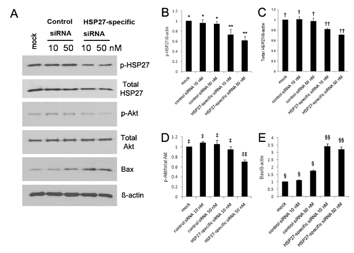
인산화 HSP27의 발현은 크게 5, 10로 증가하고, 처음 30 분 후에는 13 unwounded 인체 각막 상피 세포에 비하여 부상. 웨스턴 블롯 분석 Bax의 발현이 크게 HSP27 특정 siRNA를 형질 인체 각막 상피 세포 (그림 1A-E)에서 증가하는 반면 인산화 HSP27과 인산화 된 Akt의 발현이 모두 유의하게 감소 된 것으로 나타났습니다. 인산화 HSP27의 발현을 30 % 40 10 뉴 멕시코 % 및 HSP27 고유의 50 nm의 siRNA를 형질 감염된 세포를 각각 제어 siRNA를 형질 세포 있지만 인산화 HSP27의 발현에 비해 감소되지 않았다 (도 1A-B로 감소시켰다 ). 또한, 비 인산화 HSP27의 발현은 10 nM 내지 20 % 및 30 % 감소 하였다 siRNA를 형질 감염된 세포에 각각하지만 비 인산화 HSP27의 발현이 감소되지 않았다 (도 1a HSP27 별 및 C의 50 nM의 </ STRONG>).
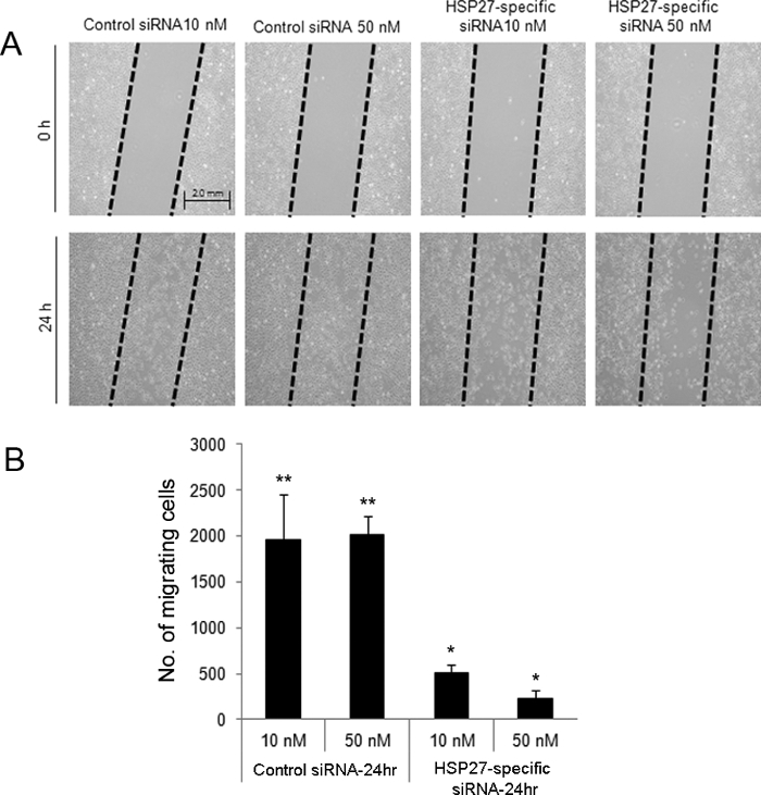
스크래치에 의한 방향 상처 분석은 상처, HSP27 특정 siRNA를 형질 세포 후 24 시간에서 10, 50 nM의 마이그레이션 (그림 2) 감소 나타내었다 것으로 나타났다. 또한, HSP27 특정 siRNA의 형질 인체 각막 상피 세포는 유동 세포 계측법에 의해 스크램블링 된 제어 siRNA를 형질 세포 (도 3)에 비해 더 많은 세포 사멸 및 괴사 성 세포 사멸을 시행 하였다.

인산화 HSP27 (p-HSP27) 비 인산화 HSP27 (비 P-HSP27), 인산화 된 Akt (Akt 인산화)는 세포 생존 마커로서 Akt의 비 인산화에 대한 항체를 사용하여도 1 웨스턴 블롯 분석 (비 P-된 Akt), BCL-2eassociated X 단백질 프로 - 세포 사멸 단백질로 (백스), 및 GAPDH (A). 비 인산화, 인산화 및 HSP27의 발현 및 제어 siRNA를 형질 감염된 세포에서 관찰되는 것과 비교 그러나 Bax의 발현은 유의하게 상기 HSP27 특이 적 siRNA를 형질 인체 각막 상피 세포 (E)의 증가 - (D B) (모두 p <0.05), 인산화 된 Akt 크게 감소 하였다. 인산화 HSP27의 발현을 30 %와 10 nm의 40 %를 각각 모의 제어와 비교 HSP27 고유의 50 nM의 siRNA를 형질 감염된 세포에 의해 감소되었지만, 인산화 HSP27의 발현은 10 nM의 감소 및 제어 siRNA를 50 nM의되지 -transfected 세포 (B). **, *; †, ††; ‡, ‡‡; §§, § : 집단 간 통계적으로 유의 한 차이 (p <0.05). 오차 막대는 표준 편차 (SD)를 나타냅니다. 이 그림의 더 큰 버전을 보려면 여기를 클릭하십시오.

그림 2. siRNA를 형질 인체 각막 상피 세포에 부상 후 세포 이동을 평가하는 방향 상처 분석을 스크래치는 유도. 스크래치 상처는 제어 및 HSP27 특정 siRNA를 형질 세포 (A)에서 만들어졌습니다. 세포는 '드래그'영역에서 제거되었습니다. 상처 후 24 시간에서, HSP27 특정 siRNA를 형질 감염된 세포를 10 내지 50nm가 10 내지 50nm 제어 siRNA를 형질 세포 (B)에 비하여 세포 이주 낮은 수치를 나타내었다. ** 및 * 그룹들 사이에 통계적으로 유의 한 차이 (p <0.05)를 나타냅니다. 데이터는 ± 표준 편차를 의미있다. 도시 이 그림의 더 큰 버전을 보려면 여기를 클릭하십시오.
 <스크램블 siRNA의 제어 및 아 넥신 V 및 PI (A 및 B)로 표지 HSP27 특정 siRNA의 형질 각막 상피 세포 (인체 각막 상피 세포)를 50 nm 인 BR />도 3 유세포. 사분면의 전체 세포의 비율은 늦게 사멸 세포 (아 넥신 V 양성과 PI 양성 세포, Q2, 오른쪽), (Q4, 오른쪽 아래, 아 넥신 V 양성과 PI-음성 세포) 초기 사멸 세포에 맞습니다 .. 및 괴사 세포 (아 넥신 V-부정과 PI 양성 세포, Q1, 왼쪽 위). HSP27 특정 siRNA를-트랜의 인체 각막 상피 세포는 siRNA를 형질 세포를 제어보다 더 많은 세포 자멸사 및 괴사 세포 사멸을 가지고 있었다. 이 그림의 더 큰 버전을 보려면 여기를 클릭하십시오.
토론
In this present study, we evaluated the potential role of HSP27 in corneal epithelial wounding using in vitro approaches. The critical steps involved siRNA transfection for HSP27 knock-down to observe the function of HSP27 in cells subjected to stress. Notably, a role for HSP27 was revealed by these experiments in epithelial cell migration and apoptosis during corneal epithelial wound healing. Unlike previous studies10 that used rat HSP27-specific siRNA to transfect vascular smooth muscle cells, we used a siRNA transfection technique to modify gene expression in human CECs to effectively knock-down HSP27-specific gene expression and study HSP27 function. Although there were differences in the target sequence that we used as well as in the cell density, final siRNA concentration, and incubation time, the protocol recommended by the manufacturer was explicitly followed. In terms of alternative methods, HSP27 knock-out mouse may be used to show if HSP27 phosphorylation involves epithelial migration and cell apoptosis. However, it is difficult to monitor the change of HSP27 phosphorylation in mouse model, because its phosphorylation occurs in very short period during epithelial wound healing.
There were several limitations to the present study. First, the in vitro environment in which we cultured human CECs certainly differed from the in vivo environment for human CECs, especially regarding cell survival. Second, the siRNA used in this study was not specific to the phosphorylated form of HSP27 as it affected the overall expression levels of HSP27, including both phosphorylated and non-phosphorylated forms.
In the future, a clinical application of these procedures would be to apply HSP27 to live human wounded corneas. We hope that the current findings will help to advance treatments of corneal epithelial tissue damage.
공개
저자는이 연구에서 언급 된 모든 자료 나 방법에는 금융 또는 소유권 관심이 없습니다.
감사의 말
이 연구는 의학, 서울, 울산 대학교 의과 대학 학생 연구 그랜트 (13 ~ 14)과 아산 생명 과학 연구소, 서울, 한국에서 보조금 (2014-464)에 의해 지원되었다.
자료
| Name | Company | Catalog Number | Comments |
| Biological safety cabinet | CHC LAB Co.Ltd, Daejeon, Republic of Korea | CHC-777A2-06 | Class II, Type A2 |
| Stealth RNAi™ siRNA | Thermo Fisher Scientific, Inc., Waltham, MA | RNAi siRNA; scrambled control-siRNA and HSP27-specific siRNA | |
| BEGMTM | Lonza, Inc., Walkersville, MD | CC-3171, CC4175 | Bronchial epithelium growth medium |
| Protease inhibitor | Sigma-Aldrich, Inc., St. Louis, MO | P8340 ,P7626 | 1 μM Pepstatin A, 1 μM Leupeptin, 0.1 μM Aprotinin |
| Bradford protein assay | Bio-Rad Laboratories, Hercules, CA | #500-0001 | Bradford protein assay |
| Nitrocellulose filters | Amersham, Little Chalfont, UK | RPN3032D | Western blotting membrane |
| Non-phosphorylated HSP27 | Abcam Inc., Cambridge, MA | ab12351 | 1:1,000 dilution (Total HSP27) |
| Phosphorylated HSP27 (Ser85) | Abcam Inc., Cambridge, MA | ab5594 | 1:1,000 dilution HSP27 was phosphorylated at Ser85 |
| Lipofectamine® RNAiMAX reagent | Invitrogen, Carlsbad, CA | 13-778-075 | Transfection reagent |
| Phosphorylated Akt (Ser473) | Cell Signaling Technology, Danvers, MA | No. 4060 | 1:1,000 dilution Akt was phosphorylated at Ser473 (cell survival marker) |
| Non-phosphorylated Akt | Cell Signaling Technology, Danvers, MA | No. 4061 | 1:1,000 dilution (Total Akt) |
| Bcl-2-associated X protein | Cell Signaling Technology, Danvers, MA | No. 4062 | 1:1,000 (anti-apoptotic protein marker) |
| GAPDH | Santa Cruz Biotechnology, Santa Cruz, CA | No. 4063 | 1:1,000 loading control marker (house keeping gene) |
| Horseradish peroxidase-conjugated goat anti-rabbit antibodies | Thermo Fisher Scientific, Inc., Waltham, MA | NCI1460KR | 1:10,000 dilution |
| OPTI-MEM | Invitrogen, Carlsbad, CA | 31985 | reduced serum medium for transfection |
| Image analysis software | Olympus, Inc., Tokyo, Japan | Image-Pro Plus 5.0 | |
| Skimed milk powder | Carl Roth GmbH + Co. KG, Karlstruhe, Germany | T145.2 | |
| Tris | Amresco LCC, Inc. Solon, OH | No-0497 | |
| Sodium Chloride | Amresco LCC, Inc. Solon, OH | No-0241 | |
| Six well culture plate | Thermo Fisher Scientific, Inc., Waltham, MA | 140675 | 35.00 mm diameter / well |
| 24-well culuture dish | Thermo Fisher Scientific, Inc., Waltham, MA | 142475 | |
| Orbital shaker | N-Bioteck, Inc., Seoul, South Korea | NB1015 | |
| Bovine serum albumin | Santa Cruz Biotechnology, Santa Cruz, CA | sc-2323 | |
| BDFACSCantoTM II | BD Biosciences, Franklin Lakes, NJ | Flow cytometry | |
| X-Ray Film | Kodak, Rochester, NY | Medical X-Ray Cassette with Green 400 Screen | |
| western blotting luminol reagent | Santa Cruz Biotechnology, Santa Cruz, CA | sc-2048 | |
| FITC Annexin V Apoptosis Detection Kit I | BD Biosciences, Franklin Lakes, NJ | 556547 |
참고문헌
- Dua, H. S., Gomes, J. A., Singh, A. Corneal epithelial wound healing. Br. J. Ophthalmol. 78 (5), 401-408 (1994).
- Estil, S., Primo, E. J., Wilson, G. Apoptosis in shed human corneal cells. Invest. Ophthalmol. Vis. Sci. 41 (11), 3360-3364 (2000).
- Guay, J., et al. Regulation of actin filament dynamics by p38 map kinase-mediated phosphorylation of heat shock protein 27. J. cell. Sci. 110, 357-368 (1997).
- Park, J. W., et al. Differential expression of heat shock protein mRNAs under in vivo glutathione depletion in the mouse retina. Neurosci. Lett. 413 (3), 260-264 (2007).
- Rane, M. J., et al. Heat shock protein 27 controls apoptosis by regulating Akt activation. J. Biol. Chem. 278 (30), 27828-27835 (2003).
- Shin, K. D., et al. Blocking tumor cell migration and invasion with biphenyl isoxazole derivative KRIBB3, a synthetic molecule that inhibits Hsp27 phosphorylation. J. Biol. Chem. 280 (50), 41439-41448 (2005).
- Jain, S., et al. Expression of phosphorylated heat shock protein 27 during corneal epithelial wound healing. Cornea. 31 (7), 820-827 (2012).
- Alekseev, O. M., Richardson, R. T., Alekseev, O., O'Rand, M. G. Analysis of gene expression profiles in HeLa cells in response to overexpression or siRNA-mediated depletion of NASP. Reprod. Biol. Endocrinol. 7, 45 (2009).
- Park, H. Y., Kim, J. H., Lee, K. M., Park, C. K. Effect of prostaglandin analogues on tear proteomics and expression of cytokines and matrix metalloproteinases in the conjunctiva and corea. Exp. Eye. Res. 94 (1), 13-21 (2012).
- Voegeli, T. S., Currie, R. W. siRNA knocks down Hsp27 and increases angiotensin II-induced phosphorylated NF-kappaB p65 levels in aortic smooth muscle cells. Inflamm. Res. 58 (6), 336-343 (2009).
- Shi, B., Isseroff, R. R. Arsenite pre-conditioning reduces UVB-induced apoptosis in corneal epithelial cells through the anti-apoptotic activity of 27 kDa heat shock protein (HSP27). J. Cell. Physiol. 206 (2), 301-308 (2006).
- Shen, E. P., et al. Comparison of corneal epitheliotrophic capacity among different human blood-derived preparations. Cornea. 30 (2), 208-214 (2011).
- Song, I. S., et al. Heat shock protein 27 phosphorylation is involved in epithelial cell apoptosis as well as epithelial migration during corneal epithelial wound healing. Exp Eye Res. 118 (1), 36-41 (2014).
재인쇄 및 허가
JoVE'article의 텍스트 или 그림을 다시 사용하시려면 허가 살펴보기
허가 살펴보기This article has been published
Video Coming Soon
Copyright © 2025 MyJoVE Corporation. 판권 소유