Method Article
의 기록 세포 내 전압 응답에 대한 전기 생리 방법
요약
Sharp microelectrodes enable accurate electrophysiological characterization of photoreceptor and visual interneuron output in living Drosophila. Here we show how to use this method to record high-quality voltage responses of individual cells to controlled light stimulation. This method is ideal for studying neural information processing in insect compound eyes.
초록
Voltage responses of insect photoreceptors and visual interneurons can be accurately recorded with conventional sharp microelectrodes. The method described here enables the investigator to measure long-lasting (from minutes to hours) high-quality intracellular responses from single Drosophila R1-R6 photoreceptors and Large Monopolar Cells (LMCs) to light stimuli. Because the recording system has low noise, it can be used to study variability among individual cells in the fly eye, and how their outputs reflect the physical properties of the visual environment. We outline all key steps in performing this technique. The basic steps in constructing an appropriate electrophysiology set-up for recording, such as design and selection of the experimental equipment are described. We also explain how to prepare for recording by making appropriate (sharp) recording and (blunt) reference electrodes. Details are given on how to fix an intact fly in a bespoke fly-holder, prepare a small window in its eye and insert a recording electrode through this hole with minimal damage. We explain how to localize the center of a cell's receptive field, dark- or light-adapt the studied cell, and to record its voltage responses to dynamic light stimuli. Finally, we describe the criteria for stable normal recordings, show characteristic high-quality voltage responses of individual cells to different light stimuli, and briefly define how to quantify their signaling performance. Many aspects of the method are technically challenging and require practice and patience to master. But once learned and optimized for the investigator's experimental objectives, it grants outstanding in vivo neurophysiological data.
서문
초파리 (Drosophila의 melanogaster의) 화합물의 눈은 신경 이미지 샘플링 및 처리를위한 광 수용체 및 interneuron 배열의 기능적 조직을 조사 할 수있는 좋은 모델 시스템이며, 동물 비전. 시스템은 가장 완벽한 배선도 1,2-을 가지며 유전자 조작 및 3-10 (높은 신호 - 대 - 잡음 비율 및 시간 해상도) 정확한 신경 활동 모니터링에 호감이다.
초파리의 눈은 함께가 머리 주위 거의 모든 방향을 커버하는 탁 트인 시야 비행을 제공 ommatidia, 개안라고 ~ 750 겉으로는 일반 렌즈 덮인 구조를 포함하는 모듈이다. 단위는 rhabdomeric 광 수용체 7,8,11 있습니다 샘플링 눈의 주요 정보를 제공합니다. 각 개안은 같은면 렌즈를 공유하지만, 일곱 가지 방향으로 정렬됩니다 팔 광 수용체 세포 (R1-R8)를 포함한다. 외부 광 수용체 R1-R6 아칸소 동안전자 파란색 - 녹색 빛에 가장 민감, 같은 방향으로 서로 지점의 상단에 거짓말 내부 세포 R7과 R8의 스펙트럼 감도, 전시 세 가지 독특한 하위 유형이 : 창백한 노란색과 지느러미 가장자리 영역 (DRA) 12 15.
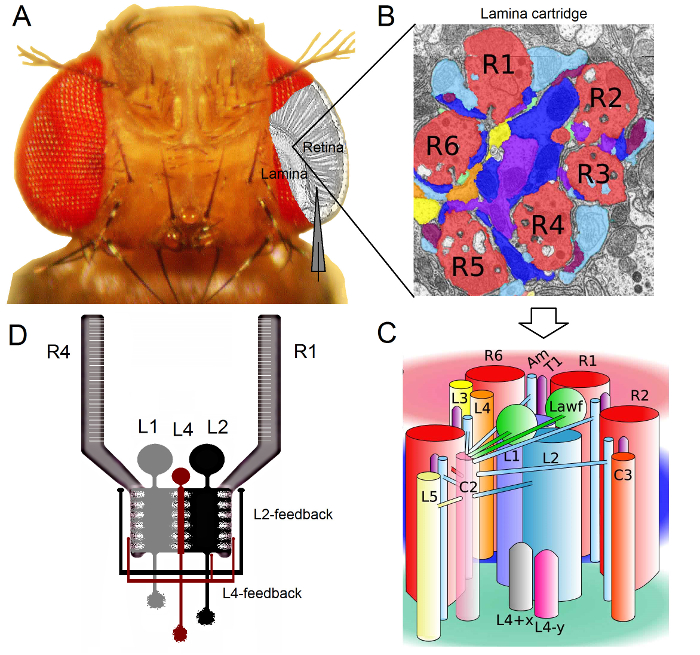
그림 1. 초파리 눈의 기능 조직. (A) 두 최초의 광학 신경, 망막과 얇은 판은 플라이 눈 안쪽 회색으로 강조 표시됩니다. 망막 R1-R6의 광 수용체와 얇은 대형 모노 폴라 셀 (중저 소득 : L1-L3)는 기존의 날카로운 미세 전극 녹음에 생체 내에서 쉽게 액세스 할 수 있습니다. 개략적 전극 망막 R1-R6에서 기록하는 정상 경로를 강조한다. 라미에 중저 소득 국가에서 기록하는 하나의 경로는 왼쪽으로 평행 전극으로 이동하는 것입니다. (B)는 얇은 판 retinotopically 기관의 행렬시각적 공간에서 특정 작은 영역에서 정보를 처리하는 신경 세포들이 즐비 각각화된 카트리지. 신경 중첩으로 인해, 다른 이웃 ommatidia, 개안로부터 6 광 수용체는 L1-L3와 무 축삭 세포 (암)에 히스타민 출력 시냅스를 형성, 동일한 라미 카트리지에 자신의 축삭 (R1-R6)를 보낼 수 있습니다. (C) R1-R6 엑손 단자와 라미 카트리지 복잡한 내부 (L4, L5, Lawf, C2, C3 및 T1 포함) 사이의 interneurons 시각 신경 정보의 확산. (D) R1-R6의 광 수용체의 축삭은 L2 및 L4 극성 세포에서 시냅스 피드백을받을 수 있습니다. 리베라 - 알바 등의 알 2에서 수정 (B)와 (C). 이 그림의 더 큰 버전을 보려면 여기를 클릭하십시오.
초파리의 눈은 신경 중첩 형 (16)이다. 이 t를 의미한다라미와 수질 : 모자는 공간에서 같은 지점에서 볼 ommatidia, 개안 이웃 일곱에 속하는 여덟 광 수용체의 신경 신호는 다음 두 neuropils 한 신경 카트리지에서 함께 풀링됩니다. 여섯 외부 광 수용체 라미에 신경 컬럼에 자신의 축삭 단자 R1-R6 프로젝트 (그림 1), R7 및 R8 세포는이 계층을 무시하고 자신이 수질 열 17-19 대응 시냅스 접촉을하는 동안. 이러한 정확한 배선은 모든 얇은 판 (도 1A-C) 그러자, 비행 초기 비전의 retinotopic 매핑에 대한 신경 기판을 생산하고 수질 열 (카트리지) 공간에서 하나의 지점을 나타냅니다.
라미 1,2,20에서와 무 축삭 세포 (암) : R1-R6의 광 수용체에서 직접 입력은 대형 모노 폴라 셀 (L1, L2 및 L3 중저 소득)에 의해 수신된다. 이 중, L1 및 L2 주요 정보 경로 (그림 1D)를 매개하는 가장 큰 세포는, WHI 있습니다채널의 응답은 온 및 오프 에지를 이동함으로써, 움직임 검출부 (21, 22)의 계산 근거를 형성한다. L2 세포 23, 24의 L1에에 전면 후면 및 전면에서 후면 : 행동 실험은 중간 반면에, 두 개의 경로가 반대 방향의 모션 인식을 용이하게 제안합니다. 연결은 또한 L4 뉴런 이웃 카트리지 (25, 26) 사이의 측 방향 연통 중요한 역할을 할 수 있다는 것을 의미한다. 상호 시냅스는 L2 및 L4 동일한에있는 세포와 두 개의 인접한 카트리지 사이에 발견되었다. 다운 스트림, 각각의 L2 셀과 세 개의 관련 L4 세포가 공통의 목표에 자신의 축삭 프로젝트, 이웃 카트리지에서 입력이 생각하는 수질에있는 TM2 신경 세포는 전후 운동 (27)의 처리를 위해 통합 될 수 있습니다. L1 신경 세포가 모두 갭 접합과 시냅스를 통해 동일한 카트리지 L2S에서 입력을받을 수 있지만, 그들은 직접 L4s 따라서 인접 라미 카트리지에 연결되어 있지 않습니다.
R1-R6의 광 수용체의 축색 돌기에 시냅틱 피드백은 L2 / L4 회로에 속하는 뉴런 만 제공 아니라 L1 경로의 1, 2 (그림 1D). 같은 카트리지 연결이 R1과 R2에 L2로부터 L4에서 R5에 선택적으로있는 동안, 모든 R1-R6의 광 수용체 중 하나의 L4 또는 둘 모두 이웃 카트리지에서 시냅스 피드백을받을 수 있습니다. 또한, R1, R2, R4 및 R5로 오전 강한 시냅스 연결이 있고, 신경교 세포는 시냅스 네트워크에 연결되어 있으므로 신경 화상 처리부 (6)에 참가할 수있다. 마지막으로, 얇은 판에서 R1-R6 및 R6 및 R7 / R8 사이의 광 수용체 이웃 연결 축삭 갭 접합은, 각 카트리지 14,20,28에서 비대칭 정보의 표현 및 처리에 기여한다.거의 그대로 초파리에서 개별 광 수용체 및 시각의 interneurons에서 세포 내 전압 녹음은 높은 신호 대 잡음 연구를 제공연결된 뉴런 사이의 빠른 신경 계산의 의미를 만들기 위해 필요한 서브 밀리 초 해상도 3,5,7-10,29,에서 다음 페이지에 계속 데이터. 100 밀리 초 해상도 - 정밀도 수준은 상당히 잡음이 있고 일반적으로 10으로 동작 전류 결상 기술에 의해 불가능하다. 전극은 매우 작고 날카로운 팁을 가지고 있기 때문에 또한,이 방법은 세포 기관에 한정되지 않고, 작은 활성 신경 구조의 직접 기록을 제공 할 수있다; 이러한 패치 클램프 전극의 훨씬 더 큰 팁에 액세스 할 수없는 중저 소득 '수지상 나무 또는 광 수용체의 축색 돌기, 등. 중요한 것은,이 방법은 또한 대부분의 패치 클램프 애플리케이션보다 덜 침습적 구조적 손상이기 때문에 적은 연구 세포의 세포 내 환경 정보 샘플링에 영향을 미친다. 따라서, 기존의 날카로운 미세 기술이 기여하고, 신경 INFOR으로, 근본적인 발견과 원래의 통찰력을 기여 계속했다적절한 시간 규모 메이션 처리; 비전 3-10 우리의 기계론의 이해를 향상시킬 수있다.
초파리 R1-R6의 광 수용체 및 중저 소득 국가에서 생체 세포 내 녹음에 Juusola 실험실에서 수행하는 방법이 문서에서는 설명합니다. 이 프로토콜은, 적절한 전기 생리학 장비를 구성하는 방법에 대해 설명 비행을 준비하고 녹음을 수행합니다. 일부 대표 데이터를 제시하고, 몇 가지 일반적인 문제와 잠재적 인 솔루션은이 방법을 사용할 때 발생 될 수있는 논의된다.
프로토콜
다음 프로토콜은 셰필드의 대학, 북경 사범 대학의 모든 동물 관리 지침을 준수합니다.
1. 시약 및 장비 준비
- 녹화 및 빛 자극 장비 설치
- 습도 조절과 공조있다 어두운 촬영 조건을 제공하는 수단을 갖추고 전기 생리 학적 실험을 수행하기위한 적어도 2.5 × 2.5 m 기록 영역을 선택한다. 모든 내에 포함, 리그 두 거위 목으로, 실체 현미경과 차가운 광원 [자극 및 기록 장치 비행] 주택 (ⅰ) 1 × 1m 진동 절연 테이블 :이 영역은 편안하게 맞게 할 수있을만큼 충분히 큰지 확인합니다 큰> 180cm 높이 패러데이 케이지; (ⅱ) 수용하는 38U 장비 랙 평면 LCD 모니터, 미세 증폭기 LED 드라이버, 필터, 온도 조절 장치, 오실로스코프 등의 필요한 전자 기기와 퍼스널 컴퓨터; 및 (iii)작은 책상 및 연구자에 대한 자입니다.
- 멀리 냉장고, 원심 분리기 및 엘리베이터 등의 전기적, 기계적 잡음 소스로부터 장비를 놓습니다. 전원에서 발생하는 전압 스파이크에서 장비의 전기 장치를 보호하기 위해 별도의 서지 보호기를 사용합니다. 이상적으로, 소음을 최소화하기 위해 자신의 무정전 전원 공급 장치 (UPS 배터리)에 장비를 연결합니다.
- 황동과 검은 색 플라스틱 (그림 2)에서 원추형 플라이 홀더를 구축합니다. 외부 림 축소에 ~ (전형적인 파리의 흉부 폭을 일치) 0.8 mm 직경 황동 장치를 통해 작은 테이퍼 구멍을 드릴.
참고 :이 구멍이 더 큰 공기 흐름 아래에서 투영되는 평균 초파리,보다 붙어 어깨 깊은 상단 가장자리에를 얻을 것입니다 수 있도록 파리 홀더의 끝을 향해 테이퍼 필요가있다. - 디자인과 비행, 기계적으로 견고하면서도 정확한 자극 및 기록 장치 (그림 3)을 구축 할 수 있습니다. 오 밖으로 구축임베디드 볼 베어링과 f를 알루미늄이나 황동 (높은 전도성 금속) 비행 준비 플랫폼 극과 주위에 카아 던 팔 시스템은, 부드럽고 정확한 X, Y 위치 결정과 광 자극의 잠금을 제공합니다.
참고 :이 통합 된 복합 디자인 그렇지 않으면 공부 세포로부터 기록 전극의 끝 부분을 제거 할 수 기계적 진동을 최소화합니다. 더 나아가 시냅스 회로 계산 9,30 평가 들면 shibire의 TS와 같은 온도에 민감한 유전자 작 제물을 사용하는 연구자있게 펠티에 소자 기반 클로즈 루프 온도 제어 시스템을 내장 할 수있다. 장치를 알루마이트 또는 검은 빛 자극 분산을 최소화하기 위해 페인트.- 방진 테이블의 브레드 보드 플라이 자극 및 기록 장치를 고정; M6 볼트에 의해, 예를 들면, 메트릭의 나사 구멍을 이용하여. 실험 기간 동안 빛 산란을 최소화하기 위해 블랙 패브릭와 함께 검은 브레드 보드를 사용하거나 커버.
- 하여 위치 될n 및 잠금 장치 카아 던 팔 시스템의 중앙에 수직으로 조정 가능한 비행 준비 플랫폼 극 (잠금 나사를 사용). 카아 던 팔에 부착 된 광원 반경 파리의 머리를 가리 키도록 플라이 준비 배치 플랫폼 극 (파리 홀더 내를, 2 단계 참조). 플라이 눈의 중심이 (0, 0) 카아 던 암의 X 축과 Y 축이 내 어느 시점에 정확한 X, 광 자극의 y 위치를 수로 파리의 교차 지점에 정확히인지 확인 시야.
참고 :이 기능은 특정 눈 위치에 개별 셀의 응답 특성을 매핑 필요하다 예를 증가 감도 나 해상도, 각각 31 보여줄 것 같은 밝은 또는 급성 영역과 같은 구조적 적응, 대한 전기 생리 증거를 검색 할 때.
- 방진 테이블에 비행 자극 뒤에 실체 현미경 및 기록 장치를 탑재 그래서이 플라이 눈의 편안한 고배율 시야를 제공.
- 광원의 듀얼 헤드 반 강성 거위 목 라이트 가이드가 비행 준비 홀더를 향해 아래로 가리키는 현미경의 상단에있는 차가운 광원을 탑재합니다. 자유 동작 개의 빔 조명 플라이 눈으로 작은 개구를 통해 구동 할 때 쉽게 기록 전극 팁을 시각화 할 수있다.
- M6 볼트 또는 자기장을 이용하여 플라이 자극 및 기록 장치의 우측에 기록 전극과 방진 테이블 헤드 스테이지 (개략 및 미세) 적합한 X, Y, Z-미세 조작기 세트 첨부 의미합니다.
참고 : Juusola 실험실에서, 다른 리그는 다른 매니퓰레이터를 갖추고 있습니다; 자세한 내용은 재료 및 시약의 표를 참조하십시오. 이들은 모두 높은 수준의 세포 내 기록을 제공한다. - 상하 조절 (F)에 기준 전극 홀더 작은 서, 3 축 micromanipulator에 마운트LY 준비 플랫폼 극. 이 비행 준비 방향을 가리 키도록 기준 전극의 방향.
- 외부 전자기 간섭을 방지하기 위해, 플라이 자극 및 기록 장치를 둘러싸고, 방진 테이블 주위 강철 패널 밖으로 독립 차광 패러데이 구축. 실험의 비행 준비를 전송하는 액세스를 제공, 케이지 개방의 전면을 남겨주세요. 소음과 빛을 보호하기 위해 전면 (접지 그들 내부에 주입 된 구리를 갖는 알루미늄 메쉬) 블랙 패브릭 커튼을 연결합니다. 광 분산을 최소화하고 진동을 방지하는 바닥 케이지의 피트를 볼트로 고정 케이지 블랙의 내부 페인트.
- BNC-케이블을 사용하여 두 개의 저역 통과 필터 (베셀 또는 이와 유사한 것)의 입력에 전압과 하이 임피던스 세포 미세 앰프의 전류 출력을 연결한다. 유사하게, AD-커넥터 BLO의 해당 채널로 필터 출력들을 연결할CKS / 데이터 수집 시스템 (DA / AD 카드)의 보드. 공급 업체의 설명서에 따라, 전문 케이블에 의한 개인 컴퓨터에 DA / AD 카드를 연결합니다.
- 개인용 컴퓨터에 선택의 데이터 수집 시스템에 적합한 수집 소프트웨어를 설치합니다. 데이터 수집 드라이버는, 퍼스널 컴퓨터의 운영 체제와 호환 가능한 것을 보장한다.
- 접지 전기적 플라이 자극 기록 장치, 패러데이 케이지 (커튼 내) 구리 메쉬 현미경 미세 조작기 감기 광원 모든 악기 38U 장비 랙 (세포 내 증폭기, 필터, 온도 제어부, PC 및 LCD 모니터 등) 설비 접지선 및 M6 링 접지 단부를 압착을 사용하여 단일 중앙 접지 지점이다. 모든 부분이 같은 지상에 있는지 테스트하기 위해 전기 멀티 미터를 사용합니다.
주 : 최상의 저잡음 기록 조건을 달성하기 위하여, 접지 구성은 일반적으로 아프로 다를다른 m 한 세트입니다.- 필요한 경우, 건물 바닥에있어서, 상기 중앙 접지 지점에 연결 및 / 또는 미세 증폭기의 중앙 접지. 실제 전기 생리 학적 실험 중에 완전히 작동 시스템을 테스트 한 결과, 레코딩의 노이즈를 최소화 할 필요 접지 구성을 변경할 수 있도록 준비.
- 신호 필터링 (모두 R1-R6와 LMC 데이터에 적합한 500 Hz에서, 설정 일반적으로 로우 패스 필터) 및 샘플링 속도 (최소 1 kHz에서) - 소프트웨어 증폭 (10X 1)을 구성합니다. 설정이 표본화 정리 (32)을 준수하는지 확인; 인 데이터 취득 할 때 예를 들어, 500 Hz에서 로우 패스 필터링 에일리어싱 효과를 최소화하기 위해 하나 이상의 kHz의 샘플링 주파수를 사용한다.
- 65 MV, 및 중저 소득 국가 (20)의 사람들 - - R1-R6의 광 수용체의 특성 전압 응답이 40이기 때문에 45 MV는, 증폭를 설정하고 디스플레이는 고해상도 sampli 수 있도록 그에 따라 확장NG 및 데이터 시각화.

그림 2. 원뿔 플라이 홀더 플라이 홀더가 두 조각으로 만들어진됩니다. 중앙 황동 유닛과 원뿔 검은 색 플라스틱 코트. 황동 기기 내부의 중앙 홀은 거의을 통해 비행을 할 수있는 작은 직경 테이퍼. 이 그림의 더 큰 버전을 보려면 여기를 클릭하십시오.

전기 생리 조작에 그림 3. 개요. 셋업은 구리 또는 함께 독립 차광 패러데이 케이지, 방진 테이블, 플라이 자극 및 기록 장치, 블랙 패브릭 커튼 포함에 대한 내부 알루미늄 메쉬 접지. 이민국trument 랙 전기적 패러데이 케이지 내부의 모든 장비와 같은 중앙 접지에 연결되어있다. 이 그림의 더 큰 버전을 보려면 여기를 클릭하십시오.
- 미소 전극을 제조
- filamented 붕규산로부터의 기준 미세 당겨 (외경 : 1.0 mm, 내경 0.6 mm) 또는 석영 유리 (외경 : 1.0 mm, 내경 0.5, 0.6 또는 0.7 mm) 피펫 풀러 기기를 사용 튜브. 짧은 점진적으로 테이퍼를 달성하려고합니다.
참고 : 피펫 풀러 프로그램의 정확한 설정이 악기 악기에서 변화; 재료 및 섭정의 표에 대한 자세한 내용. 상기 기준 전극의 선단이 즉석 제제에 삽입되기 전에 파괴 될 것이기 때문에 선단의 기공 크기는 중요하지 않다. - filamented 보로 실리케이트 (외경의 기록 미세 전극을 당겨: 1.0 mm; 내경 0.6 mm) 또는 석영 유리 (외부 직경 : 1.0 mm, 피펫 풀러 악기를 사용하여 튜브 내부 직경 0.5, 0.6 또는 0.7 mm). 미세 점진적으로 테이퍼 - (15mm 10) 긴을 달성하려고합니다.
- 기록 전극 올바른 테이퍼를 보여 광학 현미경으로 검사합니다. 성형 접착제로 유리 슬라이드에 전극을 장착하고 팁을 검사 할 40X 공기 목적을 사용합니다.
참고 : 좋은 전극이 연속 병렬 어둡고 밝은 간섭 패턴을 볼 수있는 주위의 눈에 안 띄게 작은 팁까지 부드럽게 테이퍼. 일부 풀러 설정은 팁 "나팔을"유사하기 때문에 성공적인 세포 침투를 얻을 수없는 높은 저항 전극을 생성합니다. 따라서, 전극의 육안 검사가 중요하다. - 전기 생리학 장비에 안전한 보관 및 운송에 대한 모델링 점토 (또는 유사한 성형 접착제)와 큰 페트리 접시에 수평으로 전극을 연결합니다. 있는지 확인 전극의 끝 a를공중에 항상 다시 실수를 만지기 없습니다.
- 단지 적절한 소금 용액으로 실험하기 전에-백업을 작성, 기록 전극과 기준 전극을. (예 : microloader 등) 미세 플라스틱 팁 작은 입자 필터에 연결된 작은 5 ML의 주사기를 사용합니다.
- 이 용액은 기록 전압 액간 전위의 영향을 최소화 감광체로서 실험에 3 M KCl을 가득 할 때까지 상기 기록 전극 (대형 끝에 액적 형태)를 채운다.
- 이 용액은 세포의 염화 전지에 덜 영향을 염화물 컨덕턴스 변화에 R1-R6 광 수용체에서 시냅스 입력에 응답 히스타민의 중저 소득을 조사하는, 3 M 아세트산 칼륨 및 0.5 mM의 KCl을 가진 기록 전극을 채운다. mm 단위 함유 플라이 링거액과 기준 전극을 채우 120의 NaCl, 5 KCl을 10 TES (C 6 H 15 NO 6 S), 1.5 CaCl2를 4의 MgCl 2, 30 수크로오스
5입니다.
기록 시스템에 새로 기록 인출 전극의 저항을 테스트한다.- 전극 홀더 내부의 은색 선이 균등하게 염화은으로 코팅되어 있는지 확인합니다 (보라색 회색 표시 -하지 반짝 빛) (예 : 접합 전위 드리프트)를 기록 인공물을 최소화 할 수 있습니다. 그렇지 않으면, 제대로 chloridized 와이어로 교체.
- 필요한 경우, 새로운 실버 와이어를 chloridize. 조심스럽게 이러한 색상에 밝은 실버 나타나도록 (신속 불꽃 통해 전달하여) 와이어를 청소합니다. AgCl을의 짝수 층에 증착하기 위해, 손가락으로 터치하지 마십시오. 그들은 보라색 회색 색상을 나타날 때까지 30 분 - 15 전체 강도 가정용 표백제에 전선을 만끽. 충분히 도포 될 때까지 15 초 - 대안 적으로, 10 (3 M KCl을 함유하는 용액에 대해이 긍정적 만들고 1mA 표면적 / ㎠의 속도로 그것을 통해 전류를 통과시킴으로써) 각각의 와이어를 전기 도금.
- 자신의 전극 홀더에 다시 채워진 기록과 기준 전극을 연결합니다. 수직 조정 비행 준비 플랫폼 기둥에 작은 링거액 목욕을 놓습니다. 벨소리의 용액에 전극의 끝 부분을 드라이브 및 기록 전극의 팁 저항을 측정한다.
참고 : 새 유리 튜브의 배치, 또는 때 반복을 통해 미세 전극 풀러 악기 프로그램을 최적화에서 가져온되는 전극의 저항 특성을 테스트 할 때이 단계 만 필요합니다. - 저항 측정을 수행하기 전에, 해당 측정 설정의 앰프 제조의는 사용자의 지시에 설명서를 읽어 보시기 바랍니다. 220 MΩ - 좋은 기록 전극 용 ~ (100)의 선단 저항을 갖는다.
- filamented 붕규산로부터의 기준 미세 당겨 (외경 : 1.0 mm, 내경 0.6 mm) 또는 석영 유리 (외경 : 1.0 mm, 내경 0.5, 0.6 또는 0.7 mm) 피펫 풀러 기기를 사용 튜브. 짧은 점진적으로 테이퍼를 달성하려고합니다.
2. 초파리 준비
(우화 후) 십일 된 파리와 일을 포함하는 깨끗한 플라이 튜브에 배치 - 5를 수집andard 음식. 이하 심지어 "신생아"로부터도 항공사에서 양호한 기록을 달성하는 것이 가능하다; 그러나 때문에 부드럽고 눈의 기록 전극 각막 개구부를 절단 더 어렵다.(그림 4) 서 파리 잡기 튜브와 비행 준비를 구축합니다. 이 자체 제작 도구를 함께 넣고 방법의 일반적인 아이디어를 그림 4를 참조하십시오.- 파리 잡기 튜브를하려면 50 ㎖ 플라스틱 원심 분리기 튜브의 원뿔 바닥의 끝을 잘라. 그런 다음, 삽입이 새로운 개방에 1 ml를 피펫 팁의 넓은 끝을 붙입니다.
- 마지막으로, 쉽게 비행을 통해 걸을 수있는 크기로 피펫의 작은 끝을 잘라. 2 축 회전과 다른 위치에 플라이 홀더의 잠금을 할 수있는 작은 비행 준비 단계를 조립하는 기계 워크숍을 참조하십시오.

Figur전자 4. 도구 및 고안은 비행 준비를 만들기 위해 필요. 잡기 튜브는 50 ㎖ 플라스틱 원심 관에 1 ml의 플라스틱 피펫 팁을 접착에 의해 이루어집니다 플라이. 주문품의 비행 준비 스탠드 비행을 준비하기위한 바람직한 위치에 자유 회전 및 파리 홀더의 잠금을 할 수 있습니다. 파리는 전기 왁스 히터를 사용하여, 밀랍으로 고정되어있다. 석유 젤리가 핸들에 두꺼운 정렬 머리를 연결하여 만든 작은 주걱으로 적용된다. 이 그림의 더 큰 버전을 보려면 여기를 클릭하십시오.
겨우 통해 맞게 비행을 할 수있는 1 ml를 피펫 팁의 실험 비행을 수집합니다. 플라이 튜브에, 그것에 피펫 팁, 튜브를 잡기 파리를 연결합니다. 파리 잡기에 피펫 팁으로 위쪽 (antigravitaxis)에 올라 자신의 고유 한 성향을 활용. 바람직하게는, 크기 문제로 가장 큰 여성을 선택전기 생리학에서의.
참고 : 고품질의 세포 내 녹음을위한 기회의 세포 더 큰, 비행 더 큰 및 더 나은. 작은 파리 (여성 및 남성 모두)도 우수 기록을 제공 할 수 있지만, 제조는 확인하기 어렵다. 파리가 큰 피펫 팁에 갇혀되면, 탈출 다른 파리를 중지 플라이 튜브를 닫 기억한다.피펫 팁의 더 큰 개방에 대한 유연한 플라스틱 호스와 100 ML의 주사기를 연결 - 그것은 여전히 비행과 함께.파리 홀더의 바닥에있는 구멍에, 단지를 통해 초파리를 수 있도록 확대 큰 피펫 팁에 좁은 끝을 놓고 플라이 홀더에 파리를 배출하기 위해 주사기에서 공기의 작은 볼륨을 짜내 .- 실체 현미경을 통해보고 파리의 머리는 플라이 홀더의 원추형 끝에서 돌출 될 때까지 부드럽게 더 많은 공기를 관리 할 수 있습니다. 파리 단단히 작은 OPE 그것의 가슴에서 포획되어 있는지 확인파리 홀더의 상단에 닝.
파리 홀더는 "어깨"에서 밀랍으로 비행을 고정하기 위해 왁스 히터를 사용합니다. 아직 완전히 왁스 용융 가능한 한 낮게 왁스 히터의 온도를 조정합니다.
주 : 온도가 정확하면, 왁스가 투명 나타난다. 온도가 너무 높은 왁스 "멀리 굽기"를 만든다; 너무 낮은 왁스 강성을 유지합니다. 비행을 고정 할 때 정확하고 손상 될 수 있습니다 장기 열 노출로 간단한합니다. 광중합 형 치과 용 접착제를 사용하면 해당 응용 프로그램이 너무 느립니다 여기에 사용하지 않는 것이 좋습니다.
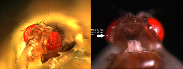
생체 실험을 위해 비행을 준비 그림 5. 초파리의 머리는 플라이 홀더에 바로 위치 및 가열와 파리의 홀더에 그 코, 오른쪽 눈과 어깨에서 고정되어, 왼쪽eswax. 좋아, 작은 개구 날카로운 면도날 에지를 사용하여, 단지 적도 멀리 백 표피에서 몇 ommatidia, 개안 위에 눈의 두꺼운 부분에서 절단된다. 각막의 조각을 조심스럽게 제거하고 구멍이 최대 건조에서 눈을 방지하기 위해 석유 젤리로 밀봉되어있다. 이 그림의 더 큰 버전을 보려면 여기를 클릭하십시오.
파리의 머리를 고정. 의 코 (그림 5)과 오른쪽 눈의 모서리에 밀랍을 적용 각막을 방지하고, 플라이 홀더 이러한 점에서 머리를 고정합니다.마이크로 칼을 생산하고 있습니다. 이 블레이드 홀더가 아닌 스테인리스 스틸 면도칼을 클램프 / 차단기 (두 평면 그립)와 날카로운 모서리의 작은 스트립 균열. 건강과 안전을 위해, (이 면도기가 금이 때 조각이 튀었 것이 매우 어렵다에도 불구하고) 눈 보호 고글을 사용합니다. 이상적으로,첨탑과 유사한 날카로운 면도날의 가장자리를 생산하고 있습니다. 이 "첨탑은"단단히 블레이드 홀더에 연결되어 있는지 확인하지만, 어떤 자기 부상을 피하기 위해 조심!기록 미세 전극을위한 통로를 제공하기 위해 단지 눈의 적도 위의 지느러미 표피에서 5 ommatidia, 개안 - 약 4에서 - 마이크로 칼을 사용하여, 파리의 좌안 몇 ommatidia, 개안의 작은 구멍 크기를 준비합니다. 높은 배율 준비를보고, 실체 현미경 아래에서 수행합니다.
참고 : 플라이 눈 프로빙에 탄성과 저항 느낌 때문에 구멍이 "첨탑"-knife 최상의 컷입니다. 절삭 기술은 매우 어려운, 그래서 비디오 데모에주의. (비행 준비 스탠드에서) 특정 방향에서 플라이 홀더를 유지하는 것은 절개를 쉽게 만들 수 있습니다. 처음에는 미세 배우기 어려운 느낄 수 있지만, 노력하면, 신경 적응은 점차 연구자의 3D-인식과 민첩성을 향상시킨다.아르 자형가져 가십시오 바로 아래에있는 망막을 노출, 절단 오프닝에서 각막 조심스럽게 작은 조각. 신속하게 바셀린 도포의 미세 머리를 사용하여 석유 젤리의 작은 덩어리와 눈의 구멍을 커버합니다.
주 : 석유 젤리 여기에 여러 역할을 제공합니다. 그것은 조직의 탈수 및 삽입 된 기록 전극을 깰 것 체액의 응고를 방지 할 수 있습니다. 그 벽내 커패시턴스를 감소 또한 부수적 코트 미세 전극. 이는 기록 시스템의 주파수 응답을 향상시키고, 기록 신경 신호의 시간 해상도 때문에 수있다. 이 광학을 흐리게으로 눈의 휴식을 통해 바셀린을 바르는하지 마십시오.
R1-R6 광수 또는 중저 소득 3. 녹화
(패러데이 케이지 또는 방진 테이블의 금속 표면을 만져 예를 들어) 미세 전극 앰프를 작동 할 때이 실수 제공에서 하나를 배제으로 항상 접지회로가 손상 될 수 있습니다 헤드 단계에 정전기를 보내고.파리 홀더가 가까운 시각 제어 선호하는 위치에 기둥에 배치 할 수 있도록 (패러데이 케이지 내부의 차가운 광원 포함)이 거위 목 라이트 가이드 (그림 6A)에 의해 위에서 비행 준비 플랫폼 극을 조명 .
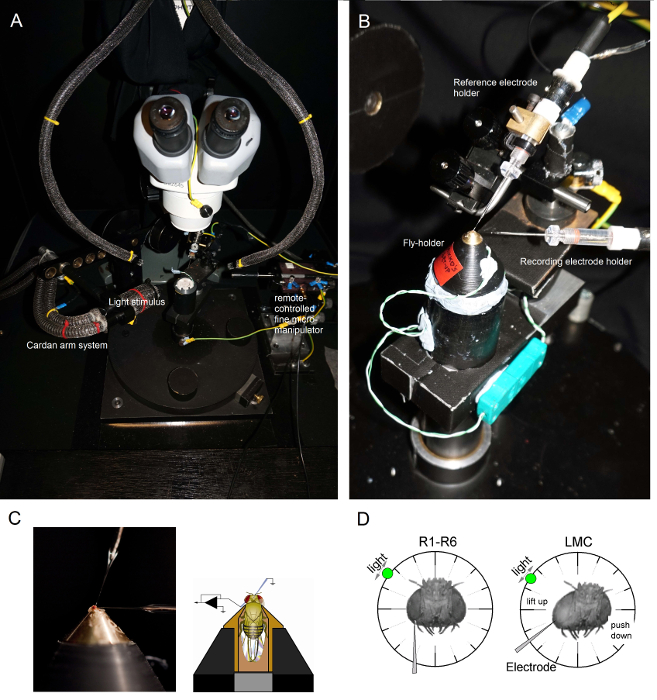
도 6 위치 플라이 홀더를 실험에 전극 (AB)이 파리의 홀더는 또한 펠티에 소자 (A : 중앙의 흰 원형 플랫폼)을 통하여 온도 제어를 제공하는 기록 플랫폼 상에 배치된다.. 카아 던 팔은 직접 눈을 가리키는 광원 (액체 또는 석영 광섬유 다발 끝)와 파리 주변 (X, Y 회전을 통해) 동일한 거리에서 빛 자극의 정확한 위치를 수 있습니다. OU의 많은에서R 리그는 빛 자극 (선형 전류 드라이버) 또는 단색으로 LED에 의해 생성됩니다. (별도의 중립적 인 밀도 필터에 의해 감쇠로) 6 로그 강도 단위 범위 - 따라서, 자신의 자극 (300) 사이에 선택된 특정 (대역 통과) 스펙트럼 내용, 수행 - 740 nm의 4 커버. 별도의 미세 조작기에 의해 제어 (C) 두 개의 미소 전극은, 플라이 헤드에 위치하는 다음 ocelli을 통해 기준 전극 (위) 왼쪽 눈에있는 작은 구멍을 통해 기록 전극 (왼쪽). 감광체 레코딩의 최대 수를 얻기 위해 (D)를, 기록 미세 구멍으로 구동되어 상기 코 - ocellus 축에 평행. 감광체에 전극 팁의 침투 및 밀봉은 자유롭게 회전 광원 셀이 주어진 광 자극에 대한 최대 전압 응답을 발생하는 위치에 고정됩니다. 공간이 시점에서는 셀의 수용 영역의 중심에있다. 구멍 난 경우, LMC 관통 더욱이 같은 전극 각도 (왼쪽)와 가까운 표피에 달성 될 수 s의. 구멍이 더 표피의 경우, 또 다른 유용한 전극 접근 각도 (오른쪽)도 표시됩니다 LMC 녹음을 얻었다. 이 그림의 더 큰 버전을 보려면 여기를 클릭하십시오.
비행 준비 플랫폼 기둥에 플라이 홀더를 (그 안에 플라이!) 마운트합니다. 파리의 왼쪽 눈에 직접 조사 (그림 6B)을 향하도록 플라이 홀더를 회전합니다.실체 현미경 (그림 6C)을 통해 준비를 관찰하면서 작은 거친 미세 조작기를 사용하여 헤드 캡슐에 파리의 occelli을 통해 조심스럽게 무딘 기준 전극을 삽입합니다. 이 플라이 뇌에 손상을 줄 수로, 너무 깊이 전극을 밀어하지 마십시오.- 대안 적으로, 흉부의 뒷면에 기준 전극을 삽입. 항상 파리가 (그 안테나 이동) 히스의 표시와 눈이 그대로 있는지 확인; 실수로 손상되지. 준비가 뽀얀보다 보이는 경우 실험에 대한 새로운 비행을 준비합니다.
석유 젤리를 통해 왼쪽 눈에 날카로운 기록 미세 전극 드라이브는 이전 준비 작은 구멍을 커버. 실체 높은 배율을 사용하여 전극의 끝 위치의 반사 패턴에 의해 3D 명백하게되도록 광 가이드 초점면을 이동한다.
참고 : 그림 6D는 파리의 머리가 광 수용체와 LMC 녹음을 위해 (기록 미세 전극이 눈에 들어가는 각도에 대하여) 다르게 약간 위치해야하는 방법을 보여줍니다. 그것을 파괴하지 않고 눈에 전극을 운전하는 것은 실험의 가장 어려운 단계입니다. 전극 팁은 작은 눈의 개폐 각막 타격을 놓치면, 그것은 일반적으로 나누기.미세 전극 앰프를 켜십시오양전극 일단 플라이 체액과 전기적으로 접촉, 제제 안에 확고하다.(패러데이 케이지 내부) 냉 광원의 전원을 끄고 전원 콘센트에서 분리합니다. 그라운드 루프는 전기 노이즈를 유도 최소화하고, 카아 던 암 시스템은 자유롭게 비행 주위를 이동할 수 있도록 멀리 거위 목 라이트 가이드를 이동하려면 중앙 땅의 플러그를 연결합니다. 비행 준비가 상대적으로 어둠에 있는지 확인하기 위해 실내 조명을 끄십시오.눈에 기록 전극 (앰프의 사용 설명서에 지시 된대로)의 저항을 측정한다. 250 MΩ - 저항이 100 인 경우에만 기록 전극을 사용합니다.
참고 : <70 MΩ 전극에 의해 고품질의 세포 내 기록을 달성하기 위해 사실상 불가능하다. 저항이 <80 MΩ 인 경우, 전극 팁이 파괴되는 것을 보인다. 이 경우에, 상기 증폭기를 전환하고, 기록 전극을 변경.- 전극 번대체 눈, 그 저항을 측정하는 앰프 스위치이다. 이 조직을 들어가는 경우에 전극의 끝 부분은 약간의 이물질에 의해 차단 될 수 있습니다. 이것은 앰프의 용량 버즈와 전류 펄스 함수를 사용하여 해결 될 수 있습니다 빠른 공진 또는 반발하여 일반적으로 분명 그.
현재 클램프 (CC) 또는 다리의 촬영 모드로 앰프를 설정합니다. 둘 다 이제 제로 오프셋 신호 (기록 전압)을 설정하여 전기적으로 연결 세포 외 공간에 쉬고, 기록 전극과 기준 전극 사이의 임의의 전압 차를 상쇄. 앰프의 디스플레이 판독하거나 오실로스코프 화면을 사용하여 신호 오프셋 변경 사항을 따르십시오.에 플라이 눈 2 ~ 3 분 기다립니다 어두운 - 적응.작은 0.1-1 미크론 단계를 눈에 점차 깊은 기록 전극의 끝 부분을 드라이브. 원격 제어 미세 조작기 또는 (B)의 X 축 피에조 스테퍼 이렇게y를 부드럽게 수동 조작의 미세 해상도 노브를 회전.기록 전극은 조직에서 진행되는로 - (10 밀리 초 1) 표시등이 깜박 간단한와 플라이 눈을 자극한다.
주 : 상기 기록 전극은 망막에 배치되고, 눈의 기능이 정상적으로 각 광 플래시 전압에 짧고 작은 방울 발생할 경우 (0.2-5 MV의 과분극을)의 전위도 (ERG)를했다. 세포 외 공간의 필드 전위의 변화는 빛에 대한 망막 세포의 집단적인 반응에 의해 발생합니다. 전극의 끝 부분은 얇은 판에 들어가면 그러나 중저 소득에 폐쇄의 ERG는 탈분극 응답을 보여주는 반전.카아 던 암 시스템을 사용하여 즉시 눈 주위의 광원을 이동하고 빛이 가장 큰 ERG 반응을 불러 일으키는 위치를 찾을 수 있습니다.
참고 :이 위치는 시각적 공간에서의 작은 영역의 표시 위치를 기록 전극의 끝 옆에 위치하는 광 수용체 (또는 중저 소득)그들의 빛 입력 샘플.기록 전극을 가진 셀을 관통.
침투가 자발적으로 발생할 수 있습니다, 또는 전극 인 경우 앞으로 마이크로 스텝 :합니다. 그것은 더 부드럽게 미세 조작기 시스템을 도청 또는 증폭기의 잡음 함수를 사용하여 촉진 될 수있다; 이러한 작업은 조직에 전극의 끝 부분을 끌어 들여. 전극이 감광체 막 impales 때, 그 세포 내 공간 기록 및 기준 전극 사이의 전압 차를 입력하면 갑자기 떨어진다 0 mV 이상 -65 ~ MV (-55 -75 MV 사이); LMC 침투 동안 반면,이 드롭 (-30과 -50 MV 사이) 일반적으로 적습니다. 이러한 전압 차이는 주어진 셀의 음극 휴지 전위를 나타낸다. 기록 전극 (그 선명도) 및 그것을 관통 세포 과정의 품질에 따라, 기록 전극의 전압 판독 세포막 같이 휴지 전위로 빠르게 서서히 안정화전극의 외층에 씰. 침투는 부분 또는 가난하지만, 전극은 일반적으로 다시 0으로 기록 된 잠재적 등반과 세포 밖으로 미끄러 져.전극이 제대로 밀봉 나타나면 안정 막 전위 (어두운 휴식 잠재력)을 보여주는 침투 세포의 수용 필드의 중심을 현지화. 빛 플래시 셀의 최대 전압 반응을 불러 일으키는 시각적 공간에 포인트를 찾으려면 카아 던 암 시스템을 사용하여, 플라이 눈 주위에 깜박이는 빛 자극을 이동합니다. 빛 자극이 직접 (에 점)에 직면 할 때 수용 필드 센터를 카아 던 팔을 잠급니다.
참고 : 어둠에서 초파리의 광 수용체는 40 밝은 빛의 펄스 응답 - 안정 LMC 녹음은 20 ~ 45 MV hyperpolarizing 응답 9,10,14을 표시하면서, 65 MV 탈분극 전압 응답 4,5. 아교 세포 침투는 <-80 MV 휴식 전위 훨씬 낮은 속도로 표시, 거의 발생하지 않을 수작은 (~ 5 MV) 광 유도 depolarizations 포화. 백색 눈 (7)과 진사와 같은 다른 눈 색소와 초파리의 광 수용체는 야생형과 비교 응답 크기를 보여줍니다.그 막 충전 중에 기록 아티팩트를 최소화하기 위해 연구 셀로 - (200 밀리 100) 전류 펄스 증폭기의 전류 클램프 (CC) 모드를 사용하여, 작은 0.1로서 NA 간단한 주입함으로써 상기 기록 전극의 커패시턴스를 보상한다.
주 :이 중요한 과정은 앰프의 사용 설명서에 상세히 설명되며, 실제 실험 전에 전기적 셀 모델로 실행한다.광 펄스와 관심의 다른 자극에 기록 전압 응답 (예 : 자연 빛의 강도의 시계열 또는 무작위 대조 패턴 등) 다양한 통계 또는 물리적 특성을 갖는. 시험, 기록 된 응답이 라이트 - 또는 어두운 적응으로 변경하는 방법 예를 들어, 용.
참고 : 하나는 리튬 정확하게 할 수 있습니다빛의 경로 4,5에 중성 밀도 필터를 추가하여 미리 선택된 강도의 연속 광에 의해 연구 된 셀을 GHT-적응. 또한, 장기간의 어두운 적응을 위해 미리 설정된 시간 동안 빛 자극을 끕니다. 때문에 기록 시스템의 기계적 안정성, 기록 전극의 품질과 제조의 intactness는 안정된 기록 조건은 종종 많은 시간 동안 지속될 수있다. 따라서, 양호한 일에, 단일 셀로부터 다른 적응시키는 상태에서 많은 양의 데이터를 수집 할 수있다. 전극이 셀의 밖으로 미끄러 때, 기록 응답은 감소하고, 평균 전압은 0에 접근하기 시작한다.전극이 접촉하고 다음 셀을 관통 할 때까지 (이는 일반적으로 신경 가까운 이웃)주의 깊게 미세 조작기의 미세 X 축 제어 기록 전극을 전진. 이러한 기동은 와줘 "는 전극을 만들 것 같은 y 축 또는 z 축을 따라 전극을 이동하지 마십시오티슈 깨닫지 못하고 있거나 남의 "옆 눈 구조 손상!
주 : 좋은 전극 및 건강 준비하여, 하나는 여러 시간에 걸쳐 동일한 즉석에서 (많은 중저 소득에서 드물게 있지만) 다수의 감광체로부터 고품질의 응답을 기록 할 수 없다; 때때로, 명확한 신호 저하없이 전체 작업 일 (> 8 시간) 이상.주기적 날짜, 유전자형, 기록 된 세포 유형으로 식별 정보 데이터 파일을 저장한다. 때문에 레코딩 세션에서 수집 될 수있는 데이터의 대량 향후 데이터 분석을 위해 실험실 책에 잘 기록 된 기록을 유지한다.
결과
초파리 눈 여기 구성된 같이 날카로운 미세 기록 방법은, 확실하게 그들 사이 4,5,7,8,10,33 신경 정보의 샘플링 처리 망막 및 점막 세포와의 통신을 정량화하는데 사용될 수있다. 다른 야생형 주식, 돌연변이 또는 유전자 조작 플라이 균주 인코딩 공부를 사용함으로써,이 방법은 그 값을 입증되었다; 뿐만 아니라 변이, 온도, 다이어트 또는 선택된 발현 3,4,6,9,10,14,30,34의 효과를 정량화 할뿐만 아니라 변경된 시각적 행동 14, 34에 대한 역학적 원인을 공개한다. 상기 방법은 또한 neuroethological 비전 연구를 실어 다른 곤충 제제 (35, 36)에 용이하게 적용될 수있다. 다음에 우리는 성공적인 응용 프로그램의 몇 가지 예를 소개.

그림 7. 전압빛 (20)의 펄스와에 과일 플라이 R1-R6 광 수용체의 응답 (25) 오 날카로운 미세 관통은 종종 매우 안정적이기 때문에 C.,에서 주어진 빛 자극에 동일한 R1-R6의 광 수용체의 전압 응답을 기록 할 수있다 온난화 또는 즉시 냉각하여 다른 주위 온도. 우리의 셋업에서, 파리 홀더 가까운 루프 펠티에 소자 기반의 온도 제어 시스템에 배치됩니다. 이 초 파리의 헤드 온도를 변경하는 우리를 할 수 있습니다. 높은 온도는 전압 응답을 가속화하고 특징적으로 (빨간색 화살표로 표시된 바와 같이) R1-R6의 광 수용체의 휴식 가능성을 낮춘다. 이 그림의 더 큰 버전을 보려면 여기를 클릭하십시오.
광 수용체 출력에 온도의 효과를 공부
와 잘 설계된 진동 절연 기록 시스템, 방법 온난화에 의해 개별 셀의 신경 출력에서 온도의 효과를 측정하거나 즉시 냉각을 위해 사용될 수있다. 주어진 예 20 및 25 O를 C (그림 7)에서 동일한 R1-R6의 광 수용체에 기록 밝은 10 밀리 긴 펄스에 전압 응답을 보여줍니다. 4,9 전에 정량으로, 온난화는 어둠 속에서 감광체의 휴식 가능성을 낮추고, 그 전압 응답을 가속화합니다.
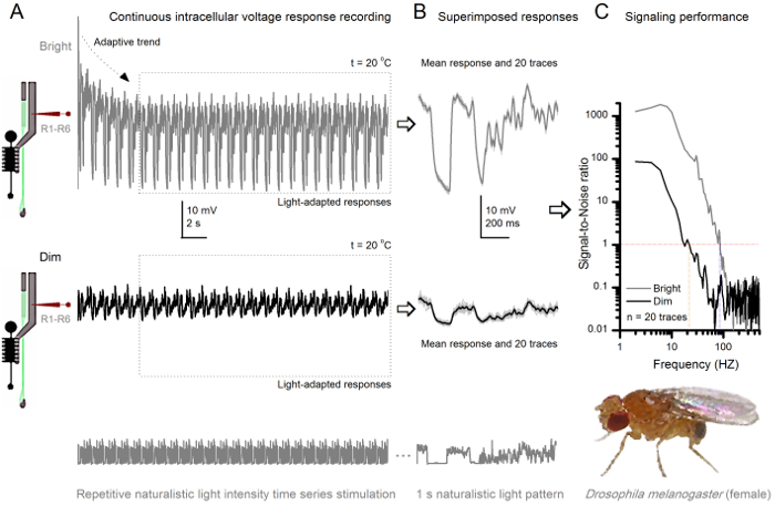
그림 8. 시그널링 성능 과일 플라이 R1-R6 광 수용체는 빛의 강도와 개선 (A) 시각 출력 (아래) 어둡게하고 밝은. (위의 10,000 배 밝은 빛) 같은 미세 전극에 의해 기록 자연 빛의 강도의 시계열을 반복20 O C.에서 같은 셀 그들은 더 많은 샘플, 단일 광자 4,5,7,8에 대한 기본 응답 (범프)를 통합하기 때문에 밝은 자극에 대한 반응이 크다. (B) 20 연속 1 초 동안 전압 응답이 중첩된다. 개별 응답 (밝은 회색) (A에서 화살표) 적응 동향 후 찍은 것은 (A에서 점선 상자) 물러했다. 대응하는 응답 수단 (신호)는 어두운 흔적이다. 신호 및 각 응답의 차이는 잡음이다. (C) 세포를 표준 방법을 사용하여 4,5,7,8 신호대 잡음비 (SNR) '시그널링 성능은 기록에 의해 정량 하였다.' 광 수용체 출력들과 함께 '(20 Hz에서 최대, 중국식 ≥1 SNR) 희미한보다 (밝은 ≥1, 최대 84 Hz의 밝은 자극에 안정적인 신호의 약 64 Hz의 넓은 범위의 SNR)'이있다ignal 잡음비 크게 개선; SNR 중국식 MAX = SNR 밝은 MAX 87 = 1868에서. 이 그림의 더 큰 버전을 보려면 여기를 클릭하십시오.
반복적 인 자극에 의해 적응과 신경 인코딩 공부
망막과 얇은 판 구조에서 상대적으로 적은 손상을 일으키는 방법의 비 침습적은 생체 내에서 자신의 근처의 자연 생리 학적 상태에서 다른 빛의 자극에 개별 세포의 신호 전달 성능을 공부에 이상적이다. 8 쇼에게 R1-의 전압 응답도 20 O C에서 희미하고 밝은 반복 자연 빛의 강도의 시계열 자극에 대한 R6의 광 수용체, 그림 9 반면, 25 오 C.에서 다른 자연 자극에 다른 R1-R6의 광 수용체와 LMC의 응답을 보여줍니다 동일한 플라이 두 예리한 마이크로 전극, 망막 하나 라미의 다른 세포에 의해 동시 기록이 가능한 30하기에는 너무 곤란하기 때문에 사전 및 시냅스 기록은 두 개의 다른 플라이에서 별도로 수행 하였다.
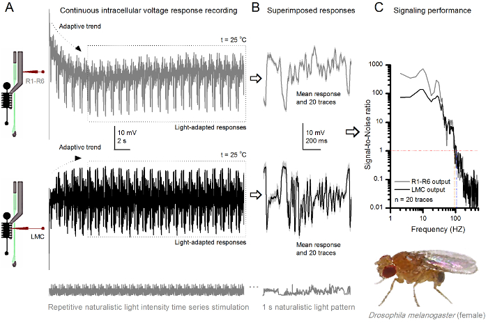
25 오 C. (A) (회색) R1-R6와 LMC (검정)에서 반복 자연주의 자극에 과일 플라이 R1-R6 광 수용체와 LMC 그림 9. 전압 응답은 다른 파리와 다른 미소 전극에 의해 기록 된 출력한다. (B) 빛에 표시된 어느 정도의 빛 적응 20 연속 전 (위) 및 개별 응답과 같은 자연 자극 패턴 시냅스 (아래) 응답을 회색어두운 트레이스로서 D 대응하는 응답 수단 (신호). 신호 및 각 응답의 차이는 잡음이다. (C) 세포의 신호대 잡음비 (SNR) '시그널링 성능은 기록에 의해 정량 하였다.' LMC 출력 '(최대 94 Hz의에 R ≥1, SNR) R1-R6 출력보다 (104 Hz에서 최대 LMC ≥1, SNR)'신뢰할 수있는 신호의 약 10 Hz의 넓은 범위를 가지고있다. 두 신호 - 대 - 잡음 비율 (SNR LMC MAX = 142 SNR R MAX = 752) 높고, 기록 노이즈가 낮고 같은 차이점은 셀 간의 실제 인코딩 차이를 반영한다. 더 큰 버전을 보려면 여기를 클릭하세요 이 그림.
자극 발병 후, 녹음 전형적인LY 빠른 크게 5 ~ 6 초 이내에 가라 동향을 적응 보여줍니다. 그때부터, 세포를 각 1 초 긴 자극 프레 젠 테이션에 매우 일관된 응답 (각 점선이 20을 둘러싸)을 생산하고 있습니다. 이 (그림 8B 및도 9b)를 중첩 할 때 응답의 반복은 분명해진다. 개별 응답은 얇은 회색 추적하고, 자신은 두꺼운 어두운 추적을 의미한다. 신경 노이즈 평균과 각 응답 4,5,9,37,38 차이 인 반면, 평균 응답은 신경 신호로한다. 주파수 영역에서 각각의 신호 - 대 - 잡음비 (도 8c 및도 9c)는 파워 스펙트럼으로 신호 및 잡음 데이터 청크를 푸리에 변환하고, 대응하는 평균 잡음 전력 스펙트럼 4 평균 신호 전력 스펙트럼을 나눔으로써 얻어졌다 5,9,37,38. 특징적으로, 최대 신호대 잡음비 O자연 자극 기록 신경 출력 (100 - 1000) >> f를 높은 값에 도달 할 수 있고, 매우 낮은 기록 노이즈 가장 안정한 제제 1,000 (예를 들면,도 8c.). 공지 사항은 또한 온난화는 세포 확장 '신호 신뢰성의 대역폭을 4 (SNR'밝은 ≥ 1); 예를 들어,도 8 및 9 개의 R1-R6s 사이의 상대적인 차이는 각각 10 헤르츠 (Hz 84 20 O C 및 94 Hz에서 25 ℃로)이다.
하나는 상기 샤논 수학 식 32를 사용하여 또는 트리플 외삽 법 (39)을 통해 응답 '엔트로피 노이즈 엔트로피 율의 차이를 산출하여 신호대 잡음비의 정보 전송의 각 셀의 속도를 추정 할 수있다. 더 많은 정보 이론적 분석에 대한 세부 사항 및 사용 제한특히이 방법으로 이전 출판물 7,8,39에 나와있다 s의.
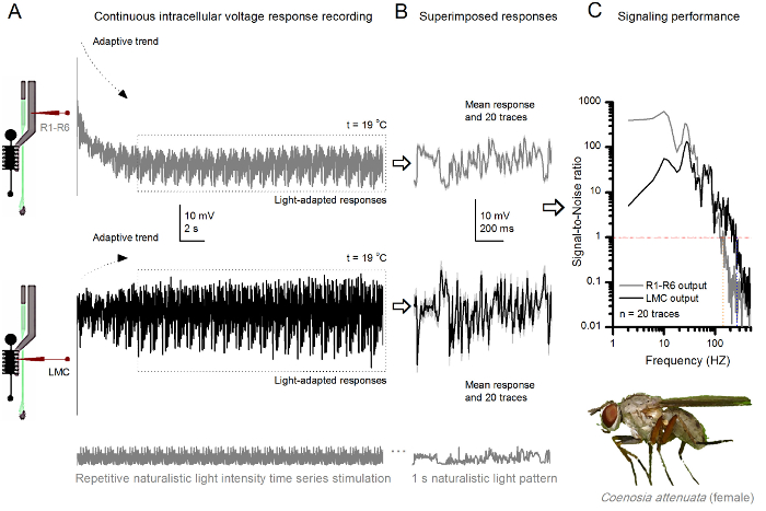
19 오 C. (A) R1-R6 (회색)와 LMC (블랙) 같은 파리에서 같은 미세 전극에 의해 기록 된 출력에서 반복 자연주의 자극에 킬러 플라이 R1-R6 광 수용체와 LMC 그림 10. 전압 응답; 전극이 눈에 진행되었을 때 이후 시냅스 처음 postsynaptically 및. 같은 자연 자극 패턴 (B) (위) 20 연속 사전 및 시냅스 (아래) 응답 (밝은 회색 추적)이 초기 적응 (A에서 점선 상자) 후 촬영 하였다. 각각의 응답에 각각의 차이는 노이즈를 제공하면서 그들의 수단은 신호 (상단에 어두운 흔적을)입니다. (C) 해당 시그나L 잡음비 (SNR)는도 8c 및도 9c에 다음과 같이 계산 하였다. LMC 출력 '(134 Hz에서 최대 R MAX ≥1 SNR) R1-R6 출력 이상 (최대 234 Hz의에 LMC MAX ≥1, SNR)'신뢰할 수있는 신호의 100 Hz의 넓은 범위에 대해이 있습니다. 두 신호 - 대 - 잡음 비율 (SNR LMC MAX = 137 SNR R MAX = 627) 높고, 동일한 미세이 녹음에 사용 된 바와 같이, 그 차이가 사전 및 시냅스 신경 출력에서 실제 차이를 반영한다. 이러한 결과는 기록 시스템이 낮은 노이즈가 있고, 분석에 미치는 영향이 한계였다. 것을 의미하는 이 그림의 더 큰 버전을 보려면 여기를 클릭하십시오.
Neuroethological 비전 연구
상기 방법은 또한 시각적 정보 처리 neuroethological 비교 연구를 허용하는 다른 곤충 7,8,35,36 종의 화합물의 눈 (도 10)의 사전 및 시냅스 전압 응답을 기록하는데 사용될 수있다. 제시된 기록 시스템의 경우, 필요한 유일한 적응은 연구 종에 대한 적절한 크기의 구멍을 가진 새로운 준비 홀더, 각이다. 이 예시적인 기록은 여성 킬러 비행 (Coenosia의 attenuata)의 출신. 이들은도 9에 대응 초파리에서 사용 된 것과 동일한 반복 광 자극에 R1-R6 수광부와 LMC 전압의 세포 내 반응을 보여 주지만, 19 ℃에서 O 이 경우, 사전 및 시냅스 데이터 모두 동일한 플라이로 기록되었다; 하나의 제 횡 박판으로 진행 (3 M KCl을 가득) 동일한 기록 전극과, 다른 하나는 이전에 입력 한 후정면 망막을 보내고. 심지어 냉각기 온도 - - 25 O C의 Coenosia 데이터의 초파리 데이터와 비교하여 빠른 반응 역학을 나타낸다 안정적인 신호 (신호 대 잡음비 >> 1)는 넓은 주파수 범위의 범위를 확대. 자연 자극의 신경 인코딩에서 이러한 기능 적응이 빠른 공중 사냥 행동을 달성하기 위해 고정밀 시공간 정보를 필요로 Coenosia의 약탈 라이프 스타일 (36)과 일치한다.
토론
We have presented the basic key steps of how to use sharp conventional microelectrodes to record intracellular responses of R1-R6 photoreceptors and LMCs in intact fly eyes. This method has been optimized, together with bespoke hardware and software tools, over the last 18 years to provide high-quality long-lasting recordings to answer a wide range of experimental questions. By investing time and resources to construct robust and precise experimental set-ups, and to produce microelectrodes with favorable electrical properties, high-quality recordings can become the norm in any laboratory working on Drosophila visual neurophysiology. Whilst well-designed recording and light stimulation systems are important for swift execution of different experimental paradigms, there are three procedural steps that are even more critical to achieving successful recordings: (i) to make the fly preparation with minimal eye damage, (ii) to pull microelectrodes with the right electrical properties, and (iii) to drive the recording electrode into the eye without breaking its tip. Ultimately, to record meaningful data, the investigator has to understand the physical basis of electrophysiology and how to fabricate suitable microelectrodes for the targeted cell-types.
Therefore, the limitations of this technique are primarily set by the patience, experience and technical ability of the investigator. Because this technique can take a long time to master for small Drosophila cells, it is advisable for trainee electrophysiologists to first practice with larger insect eyes, such as the blowfly36 or locust35, using the same rig. Once performing high-quality intracellular recordings from the larger photoreceptors and interneurons becomes routine, it is time to move on to the Drosophila eye. Another limitation of the technique concerns cellular identification. Penetrated Drosophila cells can be loaded electrophoretically with dyes, including Lucifer yellow or neurobiotin. However, because of the small tip size of the microelectrodes, electrophoresis works less efficiently than with lower resistance electrodes, such as patch-electrodes. Furthermore, the dye-filled microelectrodes characteristically have less favorable electrical properties, making it much harder to record high-quality responses with them from Drosophila photoreceptors and LMCs.
A technical problem that occurs sometimes is unstable input signal, or a complete lack of it. This is often associated with the voltage signal being either constantly drifting or higher/lower than the amplifier's recording range. On most occasions, this behavior is caused by the recording electrode being blocked (or its tip being too fine - having too high a resistance or intramural capacitance - to properly conduct fast signal changes). Although one can try to unblock the tip by buzzing the electrode capacitance, which sometimes works, often the situation is best resolved by simply changing the recording electrode. This may further require parameter adjustments in the microelectrode puller instrument to lower the tip resistance of the new electrodes. The electrode tip can also become blocked in preparations, for which it took too much time to cover the corneal hole by petroleum jelly. Prolonged air-contact can dry up the freshly exposed retinal tissue, turning its surface layer into a glue-like substance. If this is the case, the investigator typically sees a red blob of tissue stuck on the recording electrode when pulling it out of the eye. The only solution here is to make a new preparation. Petroleum jelly may provide many benefits for electrophysiological recordings: (i) it prevents the coagulation of the hemolymph that could break the electrode tip; (ii) it coats the electrode tip reducing its intramural capacitance, which lowers the electrode's time constant, and thus has the potential to improve the temporal resolution of the recorded neural signals40,41; (iii) it keeps the electrode tip clean, facilitating penetrations; and after penetration, (iv) it may even help to seal the electrode tip to the cell membrane42.
The signal can further be unstable or lost when the silver-chloride wire of the electrode-holder is broken or dechloridized; in which case just replace or rechloridize the old wire. The missing signal can also result from one (or both) of the electrode-holders not being securely connected to their jacks. However, it is extremely unusual that a piece of equipment would be malfunctioning. If signal is undetectable and all other possibilities have been exhausted, test that each part of the recording apparatus, including the headstage, amplifier, low-pass filters and AD/DA-converters, are connected properly and functioning normally. One way to achieve this is to replace each instrument with another from a rig that is known to operate normally. Alternatively, use a signal generator to check the performance of the electronic components one by one.
But perhaps the most common technical problem facing the electrophysiologist is that of recording noise. Broadly, recording noise is the observed electrical activity other than the direct neuronal response to a given stimulus. Because the fly preparation, when properly done, is very stable, the observed noise (beyond the natural variably of the responses) most often results from ground-loops in the recording equipment, or is picked up from nearby electrical devices. Such noise is typically 50/60 Hz mains hum and its harmonics; but sometimes composed of more complex waveforms. To work out the origin of the noise, remove the fly preparation holder from the set-up, connect the recording and reference electrodes through a drop of fly Ringer (or place them in a small Ringer's solution bath; see step 1.2.6) and record the signal in CC- or bridge-mode. If noise is observable on the recorded signal, this likely means that the noise is external to the fly preparation.
Another good test for identifying the origin of noise is to replace the electrode-holders with an electric cell model connected to the amplifier. In an ideally configured and grounded set-up, the recorded signal should now be practically noise-free, showing only stochastic bit-noise from the AD-converter (in the best case not even that!). If noise is still present, then recheck that all rig equipment is properly grounded. A convenient approach to detect ground-loops is to: (i) disconnect all the grounding wires from all the parts within the rig; (ii) ensure that, after doing this, every single part is actually isolated from ground, by means of an ohm-meter; (iii) connect the parts, one by one, to the central ground directly, not through any other part of the rig. Try also changing the equipment configurations. For example, sometimes moving the computer and monitor further away from the rig can reduce noise; yet at other times, moving the computer inside the equipment rack reduces noise. It is also worth unplugging nearby equipment to see if noise is reduced, or shield additional components. Furthermore, try unplugging or replacing different components of the recording equipment, especially BNC cables (which can have faulty ground connections). If only bit-noise is observed when using the cell model, the initial noise source is either the electrodes or the fly preparation itself. For example, it could be that the reference electrode is inadvertently touching a motor nerve or active muscle fibers inside the head capsule (or disturbing flight muscles in the thorax - if placed there). It is usually simplest to prepare a new fly for recording, taking care to minimize damage to the fly. But if the noise persists and is broadband, it is likely that the electrodes are suboptimal for the experiments; too sharp/fine (hence too noisy) or just wrong for the purpose; we have even seen quartz-electrodes acting as antennas - picking up faint broadcasting signals! Although iteration of the puller-instrument parameter settings to generate the just right microelectrodes for consistent high-quality recordings from specific cell-types can take a lot of effort, it is worth it. Once the recording electrodes are well-tailored for the experiments, they can provide long-lasting recordings of outstanding quality.
Sharp microelectrode recording techniques can be similarly applied to study neural information processing in multitude of preparations, including different processing layers in the insect eyes and brain43,44. Because the microelectrode tips can be made very fine, these typically damage the studied cells less than most patch-clamp applications. Importantly, the modern sample-and-hold microelectrode amplifiers enable good control of the tips' electrical properties40,45-47. Thus, when correctly applied, this technique can provide reliable data from both in vivo3,5,7-10,44 or in vitro48 preparations with high signal-to-noise ratio at sub-millisecond resolution. Such precision would be impossible with today's optical imaging techniques, which are noisier and slower. Moreover, the method can be used to characterize small cells' electrical membrane properties both in current- and voltage-clamp configurations5,29,33,36,40-42,49, providing valuable data for biophysical and empirical modeling approaches7,8,11,33,49-54 that link experiments to theory.
공개
The authors have nothing to disclose
감사의 말
The authors thank Mick Swann, Chris Askham and Martin Gautrey for their important contributions in designing and building many electrical and mechanical components of the rigs. MJ's current research is supported by the Biotechnology and Biological Sciences Research Council (BBSRC Grant: BB/M009564/1), the State Key Laboratory of Cognitive Neuroscience and Learning open research fund (China), High-End Foreign Expert Grant (China), Jane and Aatos Erkko Foundation Fellowship (Finland), and the Leverhulme Trust grant (RPG-2012-567).
자료
| Name | Company | Catalog Number | Comments |
| Stereo Zoom Microscope for making the fly preparation | Olympus | SZX12 DFPLFL1.6x PF eyepieces: WHN30x-H/22 | Capable of ~150X magnification with long working distance; bespoke heavy steel table mount stand |
| Stereomicroscope in the intracellular set-up | · Olympus | Olympus SZX7; eyepieces: WHN30x-H/22 | 30X eyepieces are needed for seeing the electrode tip reflections well when driving it through the small corneal hole into the eye |
| Nikon microscope | Nikon SMZ645; eyepieces: C-W30x/7 | ||
| Anti-vibration Table | Melles Griot | With metric M6 holes on the breadboard | Our bespoke rigs have a large hole drilled through the thick breadboard that lets in the fly preparation platform pole (houses a copper heatsink with electronics) from below |
| Newport | |||
| Micromanipulators | Narishige | Narishige NMN-21 | In our intracellular set-ups, different micromanipulator systems are used for driving the shap recording electrodes into the fly eye. All the listed manipulators are succesfully providing long-lasting stable recordings from Drosophila photoreceptors and LMCs. |
| Huxley Bertram | Huxley xyz-axis with fine manual control | ||
| Sensapex | Sensapex triple axis | ||
| Märzhäuser | Märzhäuser DC-3K with additional x-axis piezo stepper and MS 314 controller | ||
| Magnetic Stands | Any magnetic base with on/off switch will do | For example, to manage cables inside the Faraday cage | |
| Electrode Holders | Harvard Apparatus | ESP/W-F10N | |
| Silver Wire | World Precision Instruments | AGW1510 | 0.3 - 0.5 mm diameter; needs to be chloridized for the electrode holders |
| Fiber Optic Light Source | Many different, including Olympus | ||
| Fiber Optic Bundles | UltraFine Technology | To deliver the LED light stimulus to the Cardan arm system. We use both liquid and quartz light guides (range from UV to IR) | |
| Thorn Labs | |||
| Fly Cathing Tube | P80-50P 50ml Cent. Tube PP., Pack of 100 Pcs | Cut the conical bottom off from 50 ml Plastic Centrifuge Tube and glue a 1 ml pipette tip on it. | |
| Digital Acquisition System | National Instruments | ||
| Single-electrode current/voltage-clamp microelectrode amplifier | npi SEC-10LX | http://www.npielectronic.de/products/amplifiers/sec-single-electrode-clamp/sec-10lx.html | Outstanding performer! |
| Head-stage | Standard (+/- 150 nA) | For npi SEC-10LX | |
| LED light sources and drivers | 2-channel OptoLED (Cairn Research Ltd., UK) | Many of our stimulus systems are in-house built | |
| Self-designed and constructed | |||
| Acquisition and Analyses Software | Many companies to choose from | Biosyst; custom written Matlab-based system for experimental and theoretical work in the Juusola laboratory | |
| Personal Computer or Mac | Ensure that PC or Mac is compatible with data acquisition system and software | ||
| Cardan arm system | Self-designed and constructed | Providing accurate x,y,z-positioning of the light stimuli | |
| Peltier temperature control system | Self-designed and constructed | ||
| Faraday Cage | Self-constructed | Electromagnetic noise shielding | |
| Filamented Borosilicate Glass Capillaries | Outer diameter: 1 mm | ||
| Inner diameter: 0.5 - 0.7 mm | |||
| Filamented Quartz Glass Capillaries | Outer diameter: 1 mm | ||
| Inner diameter: 0.5 - 0.7 mm | |||
| Pipette Puller | Sutter Instrument Company | Model P-2000 laser Flaming/Brown Micropipette Puller | For borosilicate reference electrodes, use the preset program #11 (patch electrodes): Heat = 350; Filament = 4; Velocity 36; Delay = 200).1.2.1). For borosilicate recording electrodes, use the preset program #12 (this typically pulls good conventional sharps for photoreceptor recordings): Heat = 355; Filament = 4; Velocity 50; Delay = 225; Pull = 150. For LMC recordings, which require electrodes with finer tips, these values need to be adjusted. For pulling quartz capillaries, P-2000 manual suggests programs for fine tipped microelectrodes. These programs’ preset parameters serve as useful starting points for systematic modifications to generate electrodes with good penetration success and low recording noise. |
| Extracellular Ringer Solution for the reference electrode | Chemicals from Fisher Scientific | 10326390, NaCl 10010310, KCl 10147753, TES 10161800, CaCl2 10159872, MgCl2 10000430, sucrose | See the recipe in the protocol section |
| 3 M KCl solution for filling the filamented recording microelectrode | Salts from Fisher Scientific | 10010310, KCl | |
| Petroleum jelly | Vaselin | ||
| Non-stainless steel razor blades | |||
| Blade holder/breaker | Fine Science Tools By Dumont | 10053-09 | 9 cm |
| Blu-tack | Bostik | Alternatively, use molding clay | |
| Forceps | Fine Science Tools By Dumont | 11252-00 | #5SF (super-fine tips) |
참고문헌
- Meinertzhagen, I. A., O'Neil, S. D. Synaptic Organization of Columnar Elements in the Lamina of the Wild-Type in Drosophila-Melanogaster. J Comp Neurol. 305, 232-263 (1991).
- Rivera-Alba, M., et al. Wiring Economy and Volume Exclusion Determine Neuronal Placement in the Drosophila Brain. Curr Biol. 21, 2000-2005 (2011).
- Abou Tayoun, A. N., et al. The Drosophila SK Channel (dSK) Contributes to Photoreceptor Performance by Mediating Sensitivity Control at the First Visual Network. J Neurosci. 31, 13897-13910 (2011).
- Juusola, M., Hardie, R. C. Light adaptation in Drosphila photoreceptors: II. Rising temperature increases the bandwidth of reliable signaling. J Gen Physiol. 117, 27-41 (2001).
- Juusola, M., Hardie, R. C. Light adaptation in Drosophila photoreceptors: I. Response dynamics and signaling efficiency at 25 degrees. C. J Gen Physiol. 117, 3-25 (2001).
- Pantazis, A., et al. Distinct roles for two histamine receptors (hclA and hclB) at the Drosophila photoreceptor synapse. J Neurosci. 28, 7250-7259 (2008).
- Song, Z., Juusola, M. Refractory sampling links efficiency and costs of sensory encoding to stimulus statistics. J Neurosci. 34, 7216-7237 (2014).
- Song, Z., et al. Stochastic, Adaptive Sampling of Information by Microvilli in Fly Photoreceptors. Curr Biol. 22, 1371-1380 (2012).
- Zheng, L., et al. Feedback network controls photoreceptor output at the layer of first visual synapses in Drosophila. J Gen Physiol. 127, 495-510 (2006).
- Zheng, L., et al. Network Adaptation Improves Temporal Representation of Naturalistic Stimuli in Drosophila Eye I Dynamics. Plos One. 4, (2009).
- Hardie, R. C., Juusola, M. Phototransduction in Drosophila. Curr opin neurobiol. 34, 37-45 (2015).
- Clandinin, T. R., Zipursky, S. L. Making connections in the fly visual system. Neuron. 35, 827-841 (2002).
- Wernet, M. F., et al. Homothorax switches function of Drosophila photoreceptors from color to polarized light sensors. Cell. 115, 267-279 (2003).
- Wardill, T. J., et al. Multiple Spectral Inputs Improve Motion Discrimination in the Drosophila Visual System. Science. 336, 925-931 (2012).
- Hardie, R. C., Ottoson, D. . Progress in Sensory Physiology. 5, 1-79 (1985).
- Borst, A. Drosophila's View on Insect Vision. Curr Biol. 19, 36-47 (2009).
- Kirschfeld, K. Die Projektion Der Optischen Umwelt Auf Das Raster Der Rhabdomere Im Komplexauge Von Musca. Exp Brain Res. 3, 248-270 (1967).
- Morante, J., Desplan, C. Photoreceptor axons play hide and seek. Nat Neurosci. 8, 401-402 (2005).
- Fischbach, K. F., Hiesinger, P. R. Optic lobe development. Adv Exp Med Biol. 628, 115-136 (2008).
- Shaw, S. R. Early Visual Processing in Insects. J Exp Biol. 112, 225-251 (1984).
- Joesch, M., Schnell, B., Raghu, S. V., Reiff, D. F., Borst, A. ON and OFF pathways in Drosophila motion vision. Nature. 468, 300-304 (2010).
- Clark, D. A., Bursztyn, L., Horowitz, M. A., Schnitzer, M. J., Clandinin, T. R. Defining the Computational Structure of the Motion Detector in Drosophila. Neuron. 70, 1165-1177 (2011).
- Rister, J., et al. Dissection of the peripheral motion channel in the visual system of Drosophila melanogaster. Neuron. 56, 155-170 (2007).
- Vogt, N., Desplan, C. The first steps in Drosophila motion detection. Neuron. 56, 5-7 (2007).
- Strausfeld, N. J., Braitenberg, V. Compound Eye of Fly (Musca-Domestica) - Connections between Cartridges of Lamina Ganglionaris. Z Vergl Physiol. 70, 95-104 (1970).
- Strausfeld, N. J., Campos-Ortega, J. L4-Monopolar Neuron - Substrate for Lateral Interaction in Visual System of Fly Musca-Domestica (L). Brain Res. 59, 97-117 (1973).
- Takemura, S. Y., et al. Cholinergic Circuits Integrate Neighboring Visual Signals in a Drosophila Motion Detection Pathway. Curr Biol. 21, 2077-2084 (2011).
- Shaw, S. R., Frohlich, A., Meinertzhagen, I. A. Direct Connections between the R7/8 and R1-6 Photoreceptor Subsystems in the Dipteran Visual-System. Cell Tissue Res. 257, 295-302 (1989).
- Wolfram, V., Juusola, M. Impact of rearing conditions and short-term light exposure on signaling performance in Drosophila photoreceptors. J Neurophysiol. 92, 1918-1927 (2004).
- Nikolaev, A., et al. Network Adaptation Improves Temporal Representation of Naturalistic Stimuli in Drosophila Eye II Mechanisms. Plos One. 4, (2009).
- Land, M. F. . Compound eye structure: Matching eye to environment. Adaptive Mechanisms in the Ecology of Vision. , (1999).
- Shannon, C. E. A Mathematical Theory of Communication. At&T Tech J. 27, 379-423 (1948).
- Niven, J. E., et al. The contribution of Shaker K+ channels to the information capacity of Drosophila photoreceptors. Nature. 421, 630-634 (2003).
- Randall, A. S., et al. Speed and sensitivity of phototransduction in Drosophila depend on degree of saturation of membrane phospholipids. J Neurosci. 35, 2731-2746 (2015).
- Faivre, O., Juusola, M. Visual Coding in Locust Photoreceptors. Plos One. 3, e2173 (2008).
- Gonzalez-Bellido, P. T., Wardill, T. J., Juusola, M. Compound eyes and retinal information processing in miniature dipteran species match their specific ecological demands. P Natl Acad Sci USA. 108, 4224-4229 (2011).
- Juusola, M., Kouvalainen, E., Järvilehto, M., Weckström, M. Contrast Gain, Signal-to-Noise Ratio, and Linearity in Light-Adapted Blowfly Photoreceptors. J Gen Physiol. 104, 593-621 (1994).
- Juusola, M., Uusitalo, R. O., Weckstrom, M. Transfer of Graded Potentials at the Photoreceptor Interneuron Synapse. J Gen Physiol. 105, 117-148 (1995).
- Juusola, M., de Polavieja, G. G. The rate of information transfer of naturalistic stimulation by graded potentials. J Gen Physiol. 122, 191-206 (2003).
- Weckstrom, M., Kouvalainen, E., Juusola, M. Measurement of Cell Impedance in Frequency-Domain Using Discontinuous Current Clamp and White-Noise-Modulated Current Injection. Pflug Arch Eur J Phy. 421, 469-472 (1992).
- Juusola, M. Measuring Complex Admittance and Receptor Current by Single Electrode Voltage-Clamp. J Neurosci Meth. 53, 1-6 (1994).
- Juusola, M., Seyfarth, E. A., French, A. S. Rapid coating of glass-capillary microelectrodes for single-electrode voltage-clamp. J Neurosci Meth. 71, 199-204 (1997).
- Haag, J., Borst, A. Neural mechanism underlying complex receptive field properties of motion-sensitive interneurons. Nat Neurosci. 7, 628-634 (2004).
- Rien, D., Kern, R., Kurtz, R. Synaptic transmission of graded membrane potential changes and spikes between identified visual interneurons. Eur J Neurosci. 34, 705-716 (2011).
- Muller, A., et al. Switched single-electrode voltage-clamp amplifiers allow precise measurement of gap junction conductance. Am J Physiol-Cell Ph. 276, C980-C987 (1999).
- Polder, H. R., Swandulla, D., Konnerth, A., Lux, H. D. An Improved, High-Current Single Electrode Current Voltage Clamp System. Pflug Arch Eur J Phy. 402, R35-R35 (1984).
- Richter, D. W., Pierrefiche, O., Lalley, P. M., Polder, H. R. Voltage-clamp analysis of neurons within deep layers of the brain. J Neurosci Meth. 67, 121-131 (1996).
- Juusola, M., French, A. S. The efficiency of sensory information coding by mechanoreceptor neurons. Neuron. 18, 959-968 (1997).
- Vähäsöyrinki, M., Niven, J. E., Hardie, R. C., Weckström, M., Juusola, M. Robustness of neural coding in Drosophila photoreceptors in the absence of slow delayed rectifier K+ channels. J Neurosci. 26, 2652-2660 (2006).
- Friederich, U., Coca, D., Billings, S., Juusola, M. Data Modelling for Analysis of Adaptive Changes in Fly Photoreceptors. Lect Notes Comput Sc. 5863, 34-48 (2009).
- Asyali, M. H., Juusola, M. Use of Meixner functions in estimation of Volterra kernels of nonlinear systems with delay. Ieee T Bio-Med Eng. 52, 229-237 (2005).
- French, A. S., et al. The Dynamic Nonlinear Behavior of Fly Photoreceptors Evoked by a Wide-Range of Light Intensities. Biophys J. 65, 832-839 (1993).
- Juusola, M., French, A. S. Visual acuity for moving objects in first- and second-order neurons of the fly compound eye. J Neurophysiol. 77, 1487-1495 (1997).
- Juusola, M., Weckstrom, M., Uusitalo, R. O., Korenberg, M. J., French, A. S. Nonlinear models of the first synapse in the light-adapted fly retina. J Neurophysiol. 74, 2538-2547 (1995).
재인쇄 및 허가
JoVE'article의 텍스트 или 그림을 다시 사용하시려면 허가 살펴보기
허가 살펴보기더 많은 기사 탐색
This article has been published
Video Coming Soon
Copyright © 2025 MyJoVE Corporation. 판권 소유