Method Article
小鼠胚胎肾的解剖与培养
摘要
该方案描述了一种从小鼠胚胎中分离和培养元肾小球的方法。
摘要
该方案的目的是描述一种用于解剖,分离和培养小鼠睾丸的初步方法。
在哺乳动物肾脏发育期间,两个祖细胞组织,输尿管芽和间质间质介导并相互诱导细胞机制,最终形成肾脏的收集系统和肾单位。由于哺乳动物胚胎生长在宫内,因此观察者无法接近,已经开发出器官培养。通过这种方法,可以研究肾脏器官发生期间的上皮 - 间质相互作用和细胞行为。此外,可以研究先天性肾脏和泌尿生殖道畸形的起源。经仔细解剖后,将met ric r are are are are onto onto that that on。。。。。。。。。。。。。。。。。。。。。。。但是,必须意识到条件是人造并可能影响组织中的新陈代谢。此外,由于外植体中存在的细胞外基质和基底膜,测试物质的渗透可能受到限制。
器官文化的一个主要优点是实验者可以直接进入器官。该技术便宜,简单,并且可以进行大量的修改,例如添加生物活性物质,遗传变异体的研究以及先进成像技术的应用。
引言
The mammalian kidney is derived from two primordial structures with mesodermal origin: the tubular epithelial ureteric bud and the metanephric mesenchyme. During nephrogenesis, the ureteric bud invades the metanephric mesenchyme and branches to form the collecting system. The metanephric mesenchyme gives rise to the epithelial elements of the nephrons. These processes occur in a precisely timed and spatially coordinated manner and are initiated by reciprocal inductive mechanisms. Both tissue components communicate and affect the other's cell morphogenesis.
In the 1920s, it was Boyden who performed the in vivo obstruction of the mesonephric duct in chicken, providing the first indication of inductive interactions as separated nephric blastema fail to differentiate1. At about the same time, the first successful attempts to culture chicken nephric rudiments in a hanging drop were published. Subsequently, the organ culture was developed to study tissue interactions in mammalian organogenesis. In the 1950s, Grobstein developed a technique in which metanephric rudiments could be cultured on a filter. This technique was modified by Saxén, who placed the filter on a Trowell-type screen in a culture dish1. Over the years, many modifications and applications for organ culture have emerged. The method described here is based on Saxén's technique but is simplified, as the filters float free on the medium and the diameter of the culture well only slightly exceeds the diameter of the filter, limiting unwanted movement of the filter.
Whole-organ culture is a classical, cheap, and simple but powerful tool to investigate cellular processes and intercellular communication during organogenesis. Organ culture allows for treatment with biological agents, such as growth factors, antibodies, antisense oligonucleotides, viruses, and peptides, as well as with pharmaceutical compounds and other chemicals. Also, gene function may be studied using explants derived from genetically modified mice or using inducible gene inactivation technology, such as the Cre-loxP system. This allows for the study of genetic mutations that cause embryonic lethality prior to the development of the kidney. Organ culture can also be combined with fluorescent tagging for gene function or lineage tracing and modern imaging techniques, which enable real-time monitoring of cell behavior2.
In the specific example provided here, the effect of EphrinB2-activated Eph-receptor signaling on the branching morphology of the ureteric bud was investigated. The morphology of the EphA4/EphB2 double-knockout mice suggested several severe defects in kidney development, which were detectable as early as embryonic day 11 (E11) and involved the ureteric bud, the ureter, and the common nephric duct3. Signaling via Eph receptors requires the clustering of the ligand-receptor dimer4. To over-activate Eph signaling, the kidney rudiments from E11.5 mouse embryos were cultured in the presence of clustered recombinant EphrinB2-Fc. EphrinB2 is a known ligand for the EphA4 receptor, which is expressed in the ureteric bud tips3.
研究方案
根据瑞典法规和欧盟立法(2010/63 / EU)维护小鼠。所有程序均按照瑞典伦理委员会的准则进行(许可证C79 / 9,C248 / 11和C135 / 14)。海德堡大学涉及动物科目的程序已获得卡尔斯鲁厄大学和海德堡大学动物福利干事的批准。
1.培养试剂和材料的制备
注意:使用层流罩以尽量减少污染。
- 在解剖当天,通过在无菌磷酸盐缓冲盐水(PBS)中将其与抗人Fc山羊抗体以1:5的摩尔比混合,将人Fc单独或重组嵌合ephrin蛋白融合至人Fc。在37℃孵育1小时5 。
- 通过补充Dulbecco's Modified Eagle Medium:Nutrient来制备培养基混合物F-12(DMEM / F12)与1%青霉素/链霉素(v / v),1%谷氨酰胺(v / v),1%胰岛素转铁蛋白硒(v / v),50μg/转铁蛋白和1.5μg/ mL两性霉素。
注意:10毫升的培养基足以用于16个胚胎。 - 将最终浓度为20μg/ mL的聚簇重组ephrin溶液加入到培养基中。使用聚簇人Fc作为对照。
- 通过向无菌,未涂覆的平底聚苯乙烯4孔板的每个孔中加入500μL培养基来制备培养板。
- 使用无菌镊子,小心地将一个孔径为5μm的聚碳酸酯膜过滤器放在介质顶部。将准备的板转移到培养箱(37℃/ 5%CO 2 )。确保过滤器在上表面保持干燥并漂浮。
注意:一个过滤器足够用于两个肾脏成像。 - 使用剃刀刀片切割大约30个吸头(体积:100μL)近似值距离尖端1 mm,导致更宽的开口。将提示放回移液管尖架。
2. E11.5处的Methephric Rudiments的解剖
- 通过颈椎脱位(或使用当地道德委员会批准的方法),牺牲E11.5的定时怀孕小鼠。
- 去除子宫和胚胎,如Brown 等人 6所述 。从这一点开始,在夹层显微镜下工作。
- 使用5号制表钳,将胚胎直接切割在前肢下方。每个胚胎使用新鲜的陪替氏培养皿。去除胚胎的腿和尾巴。为了做到这一点,用一对5号制表钳抓住要去除的组织,并使用第二对的尖端切割。
- 为了去除腹侧器官体积,使用一对镊子的尖端以朝向头尾方向横向打开胚胎体壁。
- 表面切割,平行于t他背侧主动脉。使用充血的,因此可见的背侧主动脉取向。使用封闭镊子的尖端小心地将体壁的腹部与背部分开,并将胚胎翻转过来。如前面那样,以朝向头尾方向横向切割体壁,并使用背侧主动脉取向。
- 一旦腹侧体壁已经脱离背部,用一对镊子抓住它,并仔细拉扯与器官块一起去除它。
注意:甲肾上腺素通常留在背部的身体壁上。它们是椭圆形,约200μm长的结构位于后肢的水平。 - 用一对镊子握住背部侧壁,腹侧朝上。使用另一对镊子,在其末端抓住背侧主动脉,并将背侧主动脉从背侧皮瓣小心地剥离。
注意:肾脏肾脏通常保留背侧主动脉。 - 使用一对镊子,切割到肾脏,以除去主动脉。然后,使用镊子的尖端仔细分离两个肾脏成胶质细胞。
- 使用镊子的尖端小心地从肾脏中去除不需要的组织。注意不要损伤间质,因为损伤可能导致不良生长和排除外植体进一步分析。
- 使用微量移液管和10μLPBS中的大开口移液管头将肾脏成分分别转移到4孔板中的聚碳酸酯膜过滤器上。
注意:由于表面张力,外植体将被薄层介质覆盖。一个过滤器对于两个肾脏成像是足够的。- 将两个肾脏中的每一个放置在过滤器的每个相应一半的中心。将板转移到37℃/ 5%CO 2的培养箱中。将板放在孵化器中,直到metanephric rud来自下一个胚胎的图像准备放在过滤器上。
- 对每个胚胎重复步骤2.3-2.9.1。
注意:通过一些实践,每个胚胎的解剖程序将需要约5分钟。 - 在37℃/ 5%CO 2下孵育肾脏成像3天。
3.固定和染色试剂的制备
- 为了在没有钙和镁(w / v)的PBS中制备4%多聚甲醛(PFA),将PFA在60℃下在PBS中溶解,继续搅拌。冷却至室温,并使用纸过滤器去除剩余的颗粒。等分并保持在4°C即时使用或冻结长期储存。
小心:PFA有毒。戴上本地安全标准中规定的个人防护装备,并在通风橱中工作。根据制度准则废弃废物,并使用指定的废物容器。 - 为了制备透化溶液,稀释Triton X-100在不含钙和镁的PBS中至0.3%(v / v)。
- 为了制备封闭溶液,将不含钙和镁的5%(v / v)山羊血清和0.1%Triton X-100加入PBS中。加入叠氮化钠至终浓度为0.02%(w / v)。储存于4°C。
小心:叠氮化钠是有毒的。按照当地的安全环保规定处理叠氮化钠。
注意:使用抗体进行染色时,请使用来自二抗的物种的5%血清以制备阻断溶液。 - 稀释生物素化的双歧杆菌凝集素1:200在封闭溶液中。稀释Alexa488缀合的链霉抗生物素蛋白1:200在封闭溶液中。
- 为了制备即用型嵌入介质(参见材料表),向0.1M Tris-HCl(pH8.5)中的25%(w / v)甘油中加入0.1g水溶性聚乙烯醇粘膜粘附剂/ mL, 。在连续搅拌下加热至50℃1小时,冷却,并使用1调节pH至8.0-8.5M NaOH。避免较高的NaOH浓度。加入100μg/ mL的N-丙酸镓聚糖和硫柳汞,最终浓度为0.02%(w / v)。在室温下搅拌30分钟。
小心:硫柳汞是有毒的。按照当地的安全环保法规处理。- 将包埋介质填充在50mL管中,平衡转子,并以3,200 xg和室温离心10分钟。将澄清溶液倒入15 mL管中,弃去沉淀。等分并在-20°C储存数周。解冻后,将溶液保持在4°C。使用前立即温至〜30°C。
- 准备尽可能多的玻片,作为带有外植体的过滤器。为此,使用即时粘合剂在玻璃滑块的两侧上粘贴18 x 18毫米的两个小盖玻片,在其间留出约15毫米的过滤器空间。
4.固定和染色
- 培养期3次天体外 (div),从培养箱中取出板,并使用微量移液管小心地从每个孔抽取培养基。确保不要接触过滤器,并保持顶部开口朝向井壁。
- 向每个孔中加入500μL的4%PFA / PBS。过滤器将浮在固定剂上。使用微量移液器,小心地滴加PFA固定剂以淹没过滤器。在室温下孵育30分钟。
注意:对于所有以下步骤,必须注意不要从过滤器上清除外植体。将吸头的开口保持在井的墙壁上。 - 取出PFA,用500μLPBS冲洗两次,浸没过滤器。向每个孔中加入500μL透化溶液,并在室温下孵育1小时。
- 用500μLPBS冲洗两次,并向每个孔中加入500μL封闭溶液。在室温下孵育1小时。
- 用更换阻塞溶液将250μL生物素化的双歧杆菌凝集素在封闭溶液中以1:200稀释,并在4℃下孵育24小时。
- 取出Dolichorus biflorus凝集素溶液,每孔用500μLPBS冲洗两次。
- 在封闭溶液中加入250μL以1:200稀释的Alexa488缀合的链霉抗生物素蛋白,并在4℃下孵育24小时。或者,在室温下孵育2小时。
注意:当在4℃下孵育时,获得更好的信噪比。 - 每孔用500μLPBS冲洗两次。使用镊子,将过滤器转移到准备好的玻璃片上,将外植体朝上。装载约50-100μL的嵌入介质。盖上60毫米长的1.5盖玻片。允许嵌入介质溶液在黑暗中室温固化2小时。继续成像或存放幻灯片,包裹在箔中,在4°C直到准备好成像。
- 具有宽视场荧光显微镜7的图像/ sup>耦合到汞灯,并使用用于蓝色激发的反射镜单元(激发带通,460-495nm;双色镜,505nm;发射带通,510-550nm)和20X Plan-Apochromat透镜,0.75数值光圈。
结果
肾脏肾脏成纤维细胞源于怀孕的黑-65近交小鼠E11.5,并进行培养。 3天后,输尿管芽分支达5次,最终导致T型输尿管芽的分枝。拍摄每个外植体,并对片段和端点的数量进行量化,以确定分枝代数并计算每个分枝的端点数( 图1 )。 ImageJ( rd代;只有8%的处理的外植体达到第 4代,而对照外植体的比例为35%)( 图1b )。因此,每个分支的终点数和每mm 2的终点数在外植体采用聚集的EphrinB2处理,此外,三分之一的外植体在输尿管芽尖中具有不寻常的形态( 图1 ),这些结果表明EphrinB2可能具有限制作用在输尿管芽分支过程中,最有可能通过激活EphA4和EphB2正向信号传导。
对于成功的实验,重要的是,在解剖期间,肾间质不被损伤。间质的任何损伤会降低诱导电位,导致输尿管芽分叉减少或缺失,可能是偏倚的根源。 图2A中的实例显示了几乎缺失间充质的外植体。输尿管芽不超过T期。 图2B显示了一个例子,其中肾间质的损伤导致生长不良和分支不足。这两个外植体都必须排除在分析之外。

图1:E11.5元肾组织培养3代,并用聚集的EphrinB2或聚簇处理ered Human Fc作为对照。 ( A )在E11.5处切除肾脏肾脏,在生物素化的双歧杆菌凝集素和Alexa488缀合的链霉抗生物素蛋白上染色。外植体用具有460-495nm的激发带通量,505nm的二色镜,510-550nm的发射带通量和20X Plan-Apochromat镜片的宽视场荧光显微镜成像。聚合重组EphrinB2(clEphrinB2)的应用导致了输尿管芽尖的分支复杂性和畸形减少(箭头)。 ( B )左图显示了clEphrinB2处理和对照外植体中的分支代。只有8%的处理的外植体达到第 4代分支,而35%的对照外植体。每个分支的端点数(中间图)和每个区域的端点(右图)被减少(每个分支CNT的终点为2.1±0.09; clEphrinB2,1.7±0.08,P = 0.007 **;每平方毫米终点:CNT,31±0.01; clEphrinB2,27±0.02; P = 0.04 *; n = 23)数据以平均值±SEM表示,并且使用不配对的Student's t检验。刻度棒=100μm。这个数字已经从Peuckert 等人修改,2016 3 。 请点击此处查看此图的较大版本。

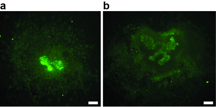
图2:在解剖期间损伤的两个E11.5肾小球肾母细胞的实例,培养3天,并用生物素化的双歧杆菌双歧杆菌凝集素和Alexa488缀合的链霉抗生物素蛋白染色。 ( A和B )间质的损伤导致生长不良或缺乏,输尿管芽分枝,取消进一步分析外植体的资格。 ( A )3周后肾肾外植体,无输尿管芽分枝。只有第一个T阶段分支是可见的。 ( B )3周后肾肾外植体,输尿管芽分叉不良。比例尺= 60μm。 请点击此处查看此图的较大版本。
讨论
该手稿描述了一种从小鼠胚胎中分离出发育中的甲基化成核细胞并培养器官基因的方法。这种方法是由Grobstein 8和Saxén9,10开发的一种标准技术,并被许多其他11,12修改和修改。该方法的成功主要取决于解剖的持续时间,随着剥离时间的延长,外植体存活和诱导电位降低。在清洁周围组织的肾脏基础时,还要注意不要损伤间质。间质间质的损伤通常是外植体生长不良的原因。然而,实践中解剖速度和精细运动技能大大提高。
所提出的方案中化学定义的介质通常用于替代含有初级细胞和体外器官培养物的含血清培养基,并含有补充有胰岛素转铁蛋白硒的DMEM和Ham's F-12的1:1(v / v)混合物,以支持外植体的生长和存活。葡萄糖和氨基酸摄取,脂肪生成和细胞内运输由胰岛素促进。硒是谷胱甘肽过氧化物酶的辅因子,起抗氧化剂的作用。铁蛋白是铁载体,有助于防止氧自由基。在胚胎肾脏培养中,向培养基中加入转铁蛋白会以剂量依赖的方式增加肾小管分化和胸苷掺入,最大效应约为50μg/ mL。 因此,人 - 全转运蛋白另外补充到培养基中,导致最终的转铁蛋白浓度为约55μg/ mL。许多协议使用了Eagle's Minimal Essential Medium(Eagle's Minimal Essential Medium),它使用更简单但化学学上较少定义的组成(MEM)或DMEM和10%血清( 即胎牛血清,FBS)也给出非常令人满意的结果12,13,14,15。然而,可能会发生不同批次的FBS之间的变化。为了避免这种变化并排除血清中存在于Eph信号中的生长因子的可能干扰,选择无血清培养基。是否使用无血清培养基的决定取决于实验设置和科学问题。当最终目标是治疗应用时,特别需要无血清培养条件。可以省略本协议中包括的抗真菌剂两性霉素B。本实施例的培养期为3天,但肾脏肾小便可培养长达10天15 。超过3天的培养基应每48小时更换一次。绑架的发展体外的初步体外重现了前管聚集体,肾囊泡和逗号和S形体的体内序列。 体外 3天后,肾小球样结构形成15,16 。在更长的文化中,由于输尿管芽继续分支,外植体区域进一步增加。培养约5天,肾单位已经分离成远端,中部和近端段16 。
尽管技术的相对简单和成本效益,允许多功能应用,但在规划实验和解释结果时,应注意一些注意事项。由于培养器官中存在细胞外基质和基底膜,外源药物和颗粒的扩散受到限制17 。此外,人造培养条件和操作可能导致代谢的变化组织的主体,细胞行为不同于体内情况 15,18。最明显的是,外植体缺血,肾小球无血管;虽然肾单位变得细分,但是分区和形成髓质和Henle的循环都缺失了14,19 。因此,胚胎肾脏培养物的应用范围限于管状结构,其分支形态和间充质 - 上皮相互作用。因此,针对肾功能的科学问题无法解决。
最近修改培养方法,其中肾组织生长在涂层玻璃上,在体积小的器官发育甚至直至皮质 - 髓质分区与Henle 15的延伸环。值得注意的是,最近,一种存储和保存生活的方法胚胎肾出版。这种方法能够在E11.5运输胚胎肾基因几天,并允许它们在以后培养。这特别关注合作20 。全肾基础文化的性质允许多种方法适应,包括先进的成像技术。为了避免在实时成像期间的干扰运动,建议使用固定的过滤器(例如transwell插图)来替换浮动。所提出的技术甚至已经扩展到含有整个泌尿生殖道的培养组织块。使用这种扩大的培养物,可以研究输尿管插入膀胱21 。
披露声明
作者没有什么可以披露的。
致谢
作者感谢Leif Oxburgh和Derek Adams慷慨分享他们的知识,Leif Oxburgh对手稿有帮助的评论,StefanWölfl和UlrikeMüller的技术支持和Saskia Schmitteckert,Julia Gobbert,Sascha Weyer和Viola Mayer在实验室。这项工作得到了"生物学家公司" (CP)发展部门的支持。
材料
| Name | Company | Catalog Number | Comments |
| DMEM/F-12 | Thermo Fisher Scientific | 21331020 | |
| Penicillin-Streptomycin (10,000 U/mL) | Thermo Fisher Scientific | 15140148 | |
| GlutaMAX Supplement | Thermo Fisher Scientific | 35050061 | |
| DPBS, calcium, magnesium | Thermo Fisher Scientific | 14040117 | use for dissection |
| holo-Transferrin human | Sigma-Aldrich | T0665 | |
| Insulin-Transferrin-Selenium (ITS -G) (100x) | Thermo Fisher Scientific | 41400045 | |
| Paraformaldehyde | Sigma-Aldrich | 158127 | |
| Amphotericin B solution | Sigma-Aldrich | A2942 | |
| Triton X-100 | Sigma-Aldrich | X100 | |
| Sodium azide | Sigma-Aldrich | S8032 | |
| Thimerosal | Sigma-Aldrich | T5125 | |
| Propyl gallate | Sigma-Aldrich | 2370 | |
| Mowiol 4-88 | Sigma-Aldrich | 81381 | |
| Glycerol | Sigma-Aldrich | G5516 | |
| Biotinylated Dolichorus Biflorus Agglutinin | Vector Laboratories | B-1035 | |
| Alexa488 conjugated Streptavidin | Jackson Immuno Research | 016-540-084 | |
| Recombinant Mouse Ephrin-B2 Fc Chimera Protein, CF | R&D Systems | 496-EB | |
| Recombinant Human IgG1 Fc, CF | R&D Systems | 110-HG-100 | |
| Goat Anti-Human IgG Fc Antibody | R&D Systems | G-102-C | |
| Phosphate buffered saline tablets | Sigma-Aldrich | P4417 | use for fixation and immunostaining |
| Dumont #5, biologie tips, INOX, 11 cm | agnthos.se | 0208-5-PS | 2 pairs of forceps are needed |
| Iris scissors, straight, 12 cm | agnthos.se | 03-320-120 | |
| Dressing Forceps, straight, delicate, 13 cm | agnthos.se | 08-032-130 | |
| Petri dishes Nunclo Delta treated | Thermo Fisher Scientific | 150679 | |
| TMTP01300 Isopore Membrane Filter, polycarbonate, Hydrophilic, 5.0 µm, 13 mm, white, plain | MerckMillipore | TMTP01300 | |
| Nunclon Multidishes 4 wells, flat bottom | Sigma-Aldrich | D6789-1CS | |
| Microscope cover glass 24 x 50 mm thickn. No.1.5H 0.17+/-0.005 mm | nordicbiolabs | 107222 | |
| Cover glasses No.1.5, 18 mm x 18 mm | nordicbiolabs | 102032 | |
| Slides ~76 x 26 x 1, 1/2-w. ground plain | nordicbiolabs | 1030418 | |
| VWR Razor Blades | VWR | 55411-055 | |
| 50 mL centrifuge tubes | Sigma-Aldrich | CLS430828 | |
| 15 mL centrifuge tubes | Sigma-Aldrich | CLS430055 | |
| Whatman prepleated qualitative filter paper, Grade 113V, creped | Sigma-Aldrich | WHA1213125 | |
| Fixed stage research mircoscope | Olympus | BX61WI | |
| Black 6 inbred mice, male, C57BL/6NTac | Taconic | B6-M | |
| Black 6 inbred mice,female, C57BL/6NTac | Taconic | B6-F | |
| Greenough Stereo Microscope | Leica | Leica S6 E |
参考文献
- Saxén, L. Organogenesis of the kidney. Developmental and Cell Biology Series. 19, Cambridge University Press. (1987).
- Lindström, N. O., et al. Integrated β-catenin, BMP, PTEN, and Notch signalling patterns the nephron. eLife. 4, e04000(2015).
- Peuckert, C., et al. Multimodal Eph/Ephrin signaling controls several phases of urogenital development. Kidney Int. 90 (2), 373-388 (2016).
- Pasquale, E. B. Eph receptor signalling casts a wide net on cell behaviour. Nat Rev Mol Cell Biol. 6 (6), 462-475 (2005).
- Bonanomi, D., et al. Ret Is a Multifunctional Coreceptor that Integrates Diffusible- and Contact-Axon Guidance Signals. Cell. 148 (2), 568-582 (2012).
- Brown, A. C., et al. Isolation and Culture of Cells from the Nephrogenic Zone of the Embryonic Mouse Kidney. J Vis Exp. (50), e2555(2011).
- Olympus Support. , Available from: http://www.olympusamerica.com/cpg_section/cpg_archived_product_details.asp?id=817 (2016).
- Grobstein, C. Inductive interaction in the development of the mouse metanephros. J Exp Zool. 130, 319-340 (1955).
- Saxén, L., Toivonen, S. Primary Embryonic Induction. , Academic Press. London. (1962).
- Saxén, L., Koskimies, O., Lahti, A., Miettinen, H., Rapola, J., Wartiovaara, J. Differentiation of kidney mesenchyme in an experimental model system. Adv Morphog. 7, 251-293 (1968).
- Dudley, A. T., Godin, R. E., Robertson, E. J. Interaction between FGF and BMP signaling pathways regulates development of metanephric mesenchyme. Genes Dev. 13, 1601-1613 (1999).
- Perälä, N., et al. Sema4C-Plexin B2 signalling modulates ureteric branching in developing kidney. Differentiation. 81 (2), 81-91 (2011).
- Thesleff, I., Ekblom, P. Role of transferrin in branching morphogenesis, growth and differentiation of the embryonic kidney. J Embryol exp Morph. 82, 147-161 (1984).
- Watanabe, T., Costantini, F. Real-time analysis of ureteric bud branching morphogenesis. Dev Biol. 271, 98-108 (2004).
- Sebinger, D. D. R., Unbekandt, M., Ganeva, V. V., Ofenbauer, A., Werner, C., Davies, J. A. A Novel, Low-Volume Method for Organ Culture of Embryonic Kidneys That Allows Development of Cortico-Medullary Anatomical Organization. PLoS One. 5 (5), e10550(2010).
- Ekblom, P., Miettinen, A., Virtanen, I., Wahlström, T., Dawnay, A., Saxén, L. In vitro segregation of the metanephric nephron. Dev Biol. 84 (1), 88-95 (1981).
- Davies, J. A., Unbekandt, M. siRNA-mediated RNA interference in embryonic kidney organ culture. Methods Mol Biol. 886, 295-303 (2012).
- Saxén, L., Lehtonen, E. Embryonic kidney in organ culture. Differentiation. 36 (1), 2-11 (1987).
- Bard, J. B. L. The development of the mouse kidney embryogenesis writ small. Curr Opin Genet Dev. 2, 589-595 (1992).
- Davies, J. A. A method for cold storage and transport of viable embryonic kidney rudiments. Kidney Int. 70 (11), 2031-2034 (2006).
- Batourina, E., et al. Distal ureter morphogenesis depends on epithelial cell remodeling mediated by vitamin A and Ret. Nat Genet. 32 (1), 109-115 (2002).
转载和许可
请求许可使用此 JoVE 文章的文本或图形
请求许可探索更多文章
This article has been published
Video Coming Soon
版权所属 © 2025 MyJoVE 公司版权所有,本公司不涉及任何医疗业务和医疗服务。