Anterograde Amnesia
Overview
Source: Laboratories of Jonas T. Kaplan and Sarah I. Gimbel—University of Southern California
Anterograde amnesia is the loss of the ability to form new memories. This can be distinguished from retrograde amnesia, which is the loss of old memories. Anterograde amnesia can result from damage to structures in the brain that are involved in the formation of new memories. Patients who have damage to the structures of the medial temporal lobe, including the hippocampus, amygdala, and the surrounding cortices, often have severe deficits in the formation of certain kinds of memories. These cases can be informative as to how memory is organized in the brain, and how different systems support different kinds of memories.
In this video, we will test a patient with medial temporal lobe damage on a series of memory tasks designed to distinguish between different forms of memory. First, we will test short-term or working memory, which is the process we use to keep information in mind temporarily. Next, we will test two different forms of long-term memory: explicit and implicit memory. Explicit memories are conscious and easy to verbalize. For example, memories of facts or episodes from our lives are explicit memories. We can easily tell someone what we ate for breakfast, or what city is the capital of France. Implicit memory involves knowledge we gain from experience but that is not easily expressible. For example, knowing how to do things, or becoming habituated to a stimulus are forms of implicit memory.
These procedures are based in part on studies of the famous patient Henry Molaison, also known by his initials H.M., who had severe anterograde amnesia as a result of a surgery for intractable epilepsy in which parts of both temporal lobes were resected.1 We will perform a test of digit span, which measures short-term memory, a test of paired-associate learning, which measures explicit or declarative memory, and mirror-drawing, a test of implicit skill learning.2
Procedure
1. Recruit participants.
- Recruit a patient with medial temporal lobe damage.
- There are several causes of medial temporal lobe damage, ranging from surgery to viral diseases and other degenerative conditions. In this study we will test a patient similar to Henry Molaison who had parts of the hippocampus and surrounding temporal lobe removed as part of a surgical treatment for epilepsy.
- In order to be able to draw conclusions about the brain structures involved in various forms of memory, perform neuroimaging to identify and describe the extent of the damaged structures.
- Recruit 15 age-matched control subjects with healthy brains and no history of neurological illness.
- Obtain informed consent from the participants.
2. Test short-term memory: Digit Span.3
- Provide instructions to the participant.
- Instruct the participant that when he or she hears a series of digits, they are to try to remember them and speak them back to the experimenter, in order, as soon as the experimenter is done listing the digits.
- Perform the Forward Digit Span Test.
- Speak a list of digits to the patient, at a rate of about 1/s. Digits are drawn from a list of randomized digits.
- Start with a sequence of four digits. Perform three trials of four digits. If the participant correctly repeats any of the three sequences of four digits without error, in proper order, move on to five-digit sequences.
- Every time the participant correctly repeats one of the series correctly, increase the length of the digit sequence. If the participant repeats none of the three series correctly, the test is done; record the maximum sequence length that the participant was able to successfully repeat.
- Perform the Reverse Digit Span Test.
- The procedure is exactly the same as the Forward Digit Span, except the participant is asked to repeat back the numbers in the reverse order. For example, if the experimenter speaks 5, 6, 1, 4, the correct response is 4, 1, 6, 5.
- Find the maximum sequence length that the participant is able to repeat using the same procedure as above.
3. Test explicit memory: Verbal Paired-associate Learning.4
- Provide instructions to the participant.
- Tell the participant that in this test, they will hear a series of word pairs, and that they should try to remember the words that go together. Tell the participant that after hearing all of the word pairs, they will be given a word and will be expected to come up with its pair. Give the participant an example: "For example, you might hear the words elephant-desk. Later, when I say elephant, I will expect you to reply desk."
- Present the study phase.
- Read the 15 word pairs to the participant, one at a time. The 15 word pairs are drawn from the list in Uttl et al., 2002.4 Read one pair of words approximately every 5 s.
- Present the testing phase.
- After the study phase is complete, wait three min. Then read the first word of each of the word pairs and prompt the participant to generate the paired word from the original list. If the participant does not respond in 10 s, or indicates that he or she does not know, that trial is considered incorrect.
4. Test implicit memory: Mirror Drawing.5
- Provide instructions to the participant.
- Tell the participant that they are to trace around the outline of the five-pointed star figure on the piece of paper in front of them, staying within the lines as much as possible. Explain that they will not be able to see their hand directly; their hand will only be seen through a mirror reflection.
- Perform the Mirror Drawing test.
- Place the participant's hand into the mirror drawing apparatus (Figure 1). This apparatus allows the participant to see their own hand only through the mirror reflection.
- Instruct the participant to trace the star shape from the starting arrow all the way around.
- Once the participant completes one drawing, wait 5 min, and begin again with a fresh star drawing.
- Complete 10 trials of the mirror drawing task.

Figure 1: The mirror drawing apparatus. This apparatus allows the participant to see his or her own hand only through the mirror reflection. The goal is to trace within the double lines of the star shape without touching the lines. This is a difficult task because visual feedback from the mirror conflicts with sensory and motor feedback from the hand.
Results
On the Digit Span Test, the patient successfully repeated a sequence of six digits in forward order, and five digits in reverse order. This level of performance shows some degree of intact short-term memory; average performance on this task for the healthy controls was seven forward, six reverse (Figure 2A). On the Verbal Paired-associate Test, the patient was not able to recall a single word pair. This demonstrates a severe deficit in the formation of explicit long term memories (Figure 2B). On the Mirror Drawing task, the amnesic patient shows fewer errors with practice, evidencing an ability to learn a motor task (Figure 2C).
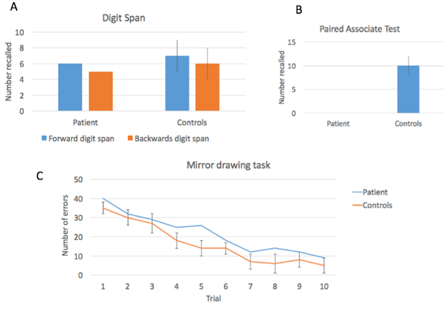
Figure 2: Performance on three memory tests. The patient showed relatively intact performance on the Digit Span Test, but was severely impaired in the Verbal Paired-associate Learning task. Performance on the motor learning task showed improvement over time.
These results demonstrate two important dissociations. The first is a dissociation between short-term memory, which requires active rehearsal to maintain, and long-term memory, the process that allows us to hold on to information without needing continual rehearsal. Patients with medial temporal lobe damage generally do not have major difficulties with short-term memory, as demonstrated by an intact digit span. However, after a few seconds, if information is not rehearsed, it is not maintained. This is demonstrated by the complete inability to retain the paired word associations in our patient. The second dissociation is between explicit and implicit memory. While this patient cannot remember the words recently seen, the Mirror Drawing test shows that they are able to learn a motor skill, a kind of learning which does not depend on medial temporal lobe structures.
Application and Summary
Cases like these have been incredibly important in the history of cognitive neuroscience for learning associations between brain structures and function. While this video has demonstrated a general effect of medial temporal lobe damage on memory function, it is important to note that a deeper understanding requires an examination of the relationship between the specifics of which structures were damaged, and memory performance. In the case of Henry Molaison, it was many years before the technology of brain imaging allowed for a clear understanding of the nature of his lesion. After he died in 2008, a post-mortem examination allowed a precise reconstruction of the lesion, showing that in addition to large portion of the hippocampus, there was also damage to the surrounding cortex and white matter fibers that carry signals into and out of the hippocampus.6
Memory loss is a consequential component of many forms of neural disease; in addition to resulting from focal brain damage, memory disturbances may result from degenerative diseases such as Alzheimer's Disease and Fronto-Temporal Dementia. Those conditions typically affect explicit long-term memories. Alternatively, motor learning, like the kind tested here, may be affected in conditions that affect the basal ganglia like Parkinson's Disease. Given the importance of memory in our lives, understanding the neural systems that underlie different forms of memory, and how memory is parcellated into different processes may also lead to techniques for improving memory performance.
References
- Scoville, W.B. & Milner, B. Loss of recent memory after bilateral hippocampal lesions. J Neurol Neurosurg Psychiatry 20, 11-21 (1957).
- Milner, B., Corkin, S. & Teuber, H.L. Further Analysis of Hippocampal Amnesic Syndrome - 14-Year Follow-up Study of Hm. Neuropsychologia 6, 215-& (1968).
- Drachman, D.A. & Arbit, J. Memory and the hippocampal complex. II. Is memory a multiple process? Arch Neurol 15, 52-61 (1966).
- Uttl, B., Graf, P. & Richter, L.K. Verbal Paired Associates tests limits on validity and reliability. Arch Clin Neuropsychol 17, 567-581 (2002).
- Milner, B. Memory and the medial temporal regions of the brain. in Biology of Memory (eds. Pribram, K.H. & Broadbent, D.E.) 29-50 (Academic Press, New York, 1970).
- Annese, J., et al. Postmortem examination of patient H.M.'s brain based on histological sectioning and digital 3D reconstruction. Nat Commun 5, 3122 (2014).
Skip to...
Videos from this collection:

Now Playing
Anterograde Amnesia
Neuropsychology
30.3K Views

The Split Brain
Neuropsychology
68.3K Views

Motor Maps
Neuropsychology
27.5K Views

Perspectives on Neuropsychology
Neuropsychology
12.0K Views

Decision-making and the Iowa Gambling Task
Neuropsychology
32.4K Views

Executive Function in Autism Spectrum Disorder
Neuropsychology
17.7K Views

Physiological Correlates of Emotion Recognition
Neuropsychology
16.2K Views

Event-related Potentials and the Oddball Task
Neuropsychology
27.5K Views

Language: The N400 in Semantic Incongruity
Neuropsychology
19.6K Views

Learning and Memory: The Remember-Know Task
Neuropsychology
17.1K Views

Measuring Grey Matter Differences with Voxel-based Morphometry: The Musical Brain
Neuropsychology
17.3K Views

Decoding Auditory Imagery with Multivoxel Pattern Analysis
Neuropsychology
6.4K Views

Visual Attention: fMRI Investigation of Object-based Attentional Control
Neuropsychology
41.7K Views

Using Diffusion Tensor Imaging in Traumatic Brain Injury
Neuropsychology
16.7K Views

Using TMS to Measure Motor Excitability During Action Observation
Neuropsychology
10.1K Views
Copyright © 2025 MyJoVE Corporation. All rights reserved