Method Article
Adaptation of Semiautomated Circulating Tumor Cell (CTC) Assays for Clinical and Preclinical Research Applications
W tym Artykule
Podsumowanie
Circulating tumor cells (CTCs) are prognostic in several metastatic cancers. This manuscript describes the gold standard CellSearch system (CSS) CTC enumeration platform and highlights common misclassification errors. In addition, two adapted protocols are described for user-defined marker characterization of CTCs and CTC enumeration in preclinical mouse models of metastasis using this technology.
Streszczenie
The majority of cancer-related deaths occur subsequent to the development of metastatic disease. This highly lethal disease stage is associated with the presence of circulating tumor cells (CTCs). These rare cells have been demonstrated to be of clinical significance in metastatic breast, prostate, and colorectal cancers. The current gold standard in clinical CTC detection and enumeration is the FDA-cleared CellSearch system (CSS). This manuscript outlines the standard protocol utilized by this platform as well as two additional adapted protocols that describe the detailed process of user-defined marker optimization for protein characterization of patient CTCs and a comparable protocol for CTC capture in very low volumes of blood, using standard CSS reagents, for studying in vivo preclinical mouse models of metastasis. In addition, differences in CTC quality between healthy donor blood spiked with cells from tissue culture versus patient blood samples are highlighted. Finally, several commonly discrepant items that can lead to CTC misclassification errors are outlined. Taken together, these protocols will provide a useful resource for users of this platform interested in preclinical and clinical research pertaining to metastasis and CTCs.
Wprowadzenie
In 2013 it is estimated that 580,350 individuals will die from cancer and that 1,660,290 new cases of this disease will be diagnosed in the United States alone1. The majority of these deaths occur subsequent to the development of metastatic disease2. The current lack of effective therapies in treating metastases and a limited understanding of the metastatic cascade makes this stage of disease highly lethal. The presence of circulating tumor cells (CTCs) within the bloodstream have been demonstrated to correlate with metastatic disease3. These cells are extremely rare and their detection is indicative of overall survival in metastatic breast4, prostate5, and colorectal6 cancer. In these patients, the presence of ≥5 (breast and prostate) or ≥3 (colorectal) CTCs in 7.5 ml of blood is indicative of poorer prognosis when compared to those patients with fewer or no detectable CTCs in the same blood volume. In addition, the change in CTC number during or after therapeutic intervention has been demonstrated to be useful as a predictor of treatment response, often sooner than currently utilized techniques7-10.
It has been estimated that, in metastatic cancer patients, CTCs occur at a frequency of approximately 1 CTC per 105-107 blood mononuclear cells and in patients with localized disease, this frequency may be even lower (~1 in 108). The rare nature of these cells can make it difficult to accurately and reliably detect and analyze CTCs11. Several methods (reviewed previously12-14) have been utilized to enrich and detect these cells by exploiting properties that differentiate them from surrounding blood components. In general, CTC enumeration is a two-part process that requires both an enrichment step and a detection step. Traditionally, enrichment steps rely on differences in physical properties of CTCs (cell size, density, deformability) or on protein marker expression (i.e. epithelial cell adhesion molecule [EpCAM], cytokeratin [CK]). Following enrichment, CTC detection can be performed in a number of different ways, the most common of which are nucleic acid-based assays and/or cytometric approaches. Each of these strategies are unique, having distinct advantages and disadvantages, however they all lack standardization; a necessity for entrance into the clinical setting. The CellSearch system (CSS) was therefore developed to provide a standardized method for the detection and enumeration of rare CTCs in human blood using fluorescence microscopy and antibody-based techniques4-6. This platform is currently considered the gold standard in CTC enumeration and is the only technique approved by the U.S. Food and Drug Administration (FDA) for use in the clinic15.
The CSS is a two component platform consisting of, (1) the CellTracks AutoPrep system (hereafter referred to as the preparation instrument), which automates the preparation of human blood samples, and (2) the CellTracks Analyzer II (hereafter referred to as the analysis instrument), which scans these samples following preparation. To distinguish CTCs from contaminating leukocytes the preparation instrument employs an antibody mediated, ferrofluid-based magnetic separation approach and differential fluorescence staining. Initially, the system labels CTCs using anti-EpCAM antibodies conjugated to iron nanoparticles. The sample is then incubated in a magnetic field, and all unlabeled cells are aspirated. Selected tumor cells are resuspended, and incubated in a differential fluorescence stain, consisting of fluorescently-labeled antibodies and a nuclear staining reagent. Finally, the sample is transferred to a magnetic cartridge, called a MagNest (hereafter referred to as the magnetic device), and scanned using the analysis instrument.
The analysis instrument is used to scan prepared samples using different fluorescence filters, each optimized to the appropriate fluorescent particle, using a 10X objective lens. CTCs are identified as cells that are bound by anti-EpCAM, anti-pan-CK-phycoerythrin (PE) (CK8, 18, and 19), and the nuclear stain 4',6-diamidino-2-phenylindole (DAPI). Conversely, contaminating leukocytes are identified as cells that are bound by anti-CD45-allophycocyanin (APC) and DAPI. Following scanning, computer-defined potential tumor cells are presented to the user. From these images, the user must employ qualitative analysis using the defined parameters and differential staining discussed above to determine which events are CTCs.
In addition to providing a standardized method for CTC enumeration, the CSS allows for molecular characterization of CTCs based on protein markers of interest. This interrogation can be performed at the single-cell level, using a fluorescein isothiocyanate (FITC) fluorescence channel not required for CTC identification16. Although this platform provides the capacity for molecular characterization, the detailed process of protocol development and optimization is not well-defined. Three commercially available markers have been developed by the manufacturer for use with the CSS, including epidermal growth factor receptor (EGFR), human epidermal growth factor receptor 2 (HER2), and insulin-like growth factor 1 receptor (IGF-1R). HER2 analysis, in combination with the CSS, has been utilized by several groups to illustrate the potential for CTC characterization to inform clinical decision-making and to potentially change existing treatment guidelines. For example, Fehm et al.17 demonstrated that approximately one third of breast cancer patients with HER2-primary tumors had HER2+ CTCs. In addition, Liu et al.18 recently reported that up to 50% of patients with HER+ metastatic breast cancer did not have HER2+ CTCs. Herceptin, a HER2 receptor interfering monoclonal antibody demonstrated to greatly benefit patients whose tumors express sufficient levels of HER2, is a commonly utilized treatment for patients with HER2+ primary tumors19-21. However, these studies suggest that Herceptin may be being sub-optimally utilized and that CTC characterization may aid in predicting treatment response. Ultimately, CTC characterization may have the potential to improve personalized care.
CTC research is unique in that it has largely utilized a bedside-to-benchtop approach. This method, unlike benchtop-to-bedside research, which can often take years to impact patient care, has allowed CTCs quick entry into the clinical setting. However, physicians are hesitant to use results from CTC analysis in patient treatment decision-making due to a lack of understanding of their underlying biology. Therefore appropriate preclinical mouse models of metastasis and complementary CTC analysis techniques must be utilized in order to investigate these outstanding questions. In general, there are two types of preclinical models used to study the metastatic cascade, (1) spontaneous metastasis models, which allow for the study of all the steps in the metastatic cascade, and (2) experimental metastasis models, which only allow for the study of later steps in the metastatic process such as extravasation and secondary tumor formation22. Spontaneous metastasis models, involve tumor cell injections into appropriate orthotopic locations (e.g. injection of prostate cancer cells into the prostate gland for the study of prostate cancer). Cells are then given time to form primary tumors and spontaneously metastasize to secondary sites such as the bone, lung, and lymph nodes. In contrast, experimental metastasis models involve direct injection of tumor cells into the bloodstream (e.g. via tail vein or intracardiac injection to target cells to specific locations) and therefore skip the initial steps of intravasation and dissemination to secondary organs22. Thus far the majority of CTC analysis in in vivo model systems has been performed using either cytometry-based23 or adapted human-based CTC techniques (e.g. AdnaTest)24. Although useful, none of these techniques adequately reflect CTC enumeration using the gold standard CSS. Based on the clinical approval, standardized nature, and widespread usage of the CSS, the development of a CTC capture and detection technique for in vivo modeling that utilizes equivalent sample preparation, processing, and identification criteria would be advantageous as results would be comparable to those obtained from patient samples. However, due to the volume requirements of the preparation instrument it is not possible to process small volumes of blood using this automated platform. In addition, previous work by Eliane et al.25 has demonstrated that contamination of samples with mouse epithelial cells (which also meet the standard CTC definition [EpCAM+CK+DAPI+CD45-]) can lead to misclassification of mouse squamous epithelial cells as CTCs. To address these issues an adapted technique that allows the utilization of the CSS CTC kit reagents combined with a manual isolation procedure was developed. The addition of a FITC labeled human leukocyte antigen (HLA) antibody to the assay allows human tumor cells to be distinguished from mouse squamous epithelial cells.
This manuscript describes the standard, commercially developed and optimized CSS protocol for processing patient blood samples and common pitfalls that may be encountered, including discrepant items that can lead to CTC misclassification errors. In addition, customization of the CSS assay to examine user-defined protein characteristics of captured CTCs and a comparable adapted CSS technique that allows for the enrichment and detection of CTCs from small volumes of blood in preclinical mouse models of metastasis are described.
Protokół
All human studies described in this manuscript were carried out under protocols approved by Western University's Human Research Ethics Board. All Animal studies were conducted in accordance with the recommendations of the Canadian Council on Animal Care, under protocols approved by the Western University Animal Use Subcommittee.
1. Standard CTC Enumeration from Patient Blood Samples Using the CSS
1. Human Blood Sample Collection and Preparation for Processing on the Preparation Instrument
- Using standard aseptic phlebotomy techniques, draw a minimum of 8.0 ml of human blood into a 10 ml CellSave tube (hereafter referred to as the CTC preservative tube), which contains ethylene diaminetetraacetic acid (EDTA) and a proprietary cellular preservative. Invert the tube 5x to prevent blood from clotting. Samples may be processed immediately or stored at room temperature for up to 96 hr.
- Remove CSS reagents from the fridge and allow them to warm to room temperature before using.
- Using a disposable 10 ml pipette and automated pipettor, collect 7.5 ml of blood from the CTC preservative tube and slowly dispense blood into an appropriately labeled preparation instrument processing tube.
- Add 6.5 ml of dilution buffer to each sample. Mix by inverting sample 5x. Centrifuge sample at 800 x g for 10 min with the brake in the "off" position. Follow the on-screen instructions on the preparation instrument to load all patient samples onto the system for processing. Samples must be processed within 1 hr of preparation.
2. Control Preparation for Processing on the Preparation Instrument
- Gently vortex the control vial and invert 5x to mix.
- Carefully remove the cap from the control vial and place an inverted preparation instrument processing tube on top of the uncapped vial. In one swift motion, invert the control vial, and pour the contents into the processing tube. While inverted, gently flick the sides of the control vial to release any remaining contents.
- Carefully remove the inverted control vial from the processing tube, ensuring that no liquid is lost and place aside. Using a 1,000 μl pipette, collect any remaining contents from the vial and lid and gently pipette into the processing tube.
- Follow the on-screen instructions on the preparation instrument to load the control onto the system for processing.
3. Sample Scanning on the Analysis Instrument
- Follow the on-screen instructions on the preparation instrument to unload all samples from the system. Loosely cap each magnetic device cartridge and tap the magnetic device using hands or lab bench to release any bubbles that are stuck to the edges of the cartridge. Once all the bubbles have been removed, firmly cap the cartridge, lay the magnetic device flat, and incubate in the dark for at least 20 min at room temperature. Samples must be scanned within 24 hr of preparation.
- Turn on the analysis instrument and initialize the lamp. Once warmed (~15 min), load the system verification cartridge onto the analysis instrument and select the QC Test tab. Follow the on-screen instructions to perform the necessary quality control measures.
- Load a sample onto the analysis instrument and select the Patient Test tab. All saved information from the preparation instrument will be displayed. Click Start to initialize sample scanning. The system will perform a coarse focus and edge detection on the magnetic device cartridge.
- Adjust all edges as necessary using the directional keys. Select Accept. The system will perform a fine focus and begin sample scanning.
- Following control scanning the results should be validated using the defined criteria for cells spiked at high (CK+DAPI+CD45-APC+) and low (CK+DAPI+CD45-FITC+) concentrations. Following patient sample scanning the results should be reviewed for captured CTCs using the defined CTC criteria (CK+DAPI+CD45-).
2. CTC Characterization for User-defined Markers using the CSS
1. Preparation of User-defined Markers and Instrument Initialization
- Dilute the antibody of interest using Bond Primary Antibody Diluent to the desired concentration in a marker reagent cup using the following formula, where the working concentration is the concentration of the antibody after addition to the sample and the stock concentration is the concentration of antibody in the reagent cup. For multiple samples, adjust the antibody volumes as described in Table 1. Place the marker reagent cup into position 1 in the reagent cartridge and load the cartridge onto CSS.
Stock Concentration = (Working Concentration x 850 μl) / 150 μl
- Collect blood, prepare samples, and load the preparation instrument as described in the above Standard CTC Enumeration from Patient Blood Samples using the CSS protocol. To enable custom marker addition, select User Defined Assay when prompted by the preparation instrument. Input the marker name and select Save. As samples are loaded onto the instrument, the operator will be prompted to indicate which should receive custom marker by selecting Yes or No as necessary.
2. Sample Scanning of User-Defined Markers on the Analysis Instrument
- Turn on the analysis instrument, initialize the lamp, and perform quality control and system verification as described in Section 3.2 of the Standard CTC Enumeration from Patient Blood Samples using the CSS protocol.
- Load a sample onto the analysis instrument and select the Setup tab. To initialize the FITC channel, select CellSearch CTC as the Kit ID under the Test Protocols section. From this menu, select CTC Research, click the Edit button and set the exposure time as desired. It is recommended that an exposure time of 1.0 sec not be exceeded when using the CSS CTC kit as this can increase bleed-through into other fluorescent channels utilized for CTC identification.
3. Adaptation of the Standard CSSProtocol for use in Preclinical Mouse Models
**Adapted from Veridex Mouse/Rat CellCapture Kit (no longer commercially available)
1. Mouse Blood Collection and Storage
- Prior to blood collection, run ~30 μl of 0.5 M EDTA back and forth through a 22 G needle, leaving a small amount of EDTA in the hub.
- Collect a minimum of 50 μl of mouse blood from mice previously injected with human tumor cells via orthotopic, tail vein, or intracardiac routes. Collect blood from the saphenous vein (for serial CTC analysis) or by cardiac puncture (for terminal CTC analysis). Remove needle and dispense blood into a 1 ml EDTA microtainer blood collection tube. Mix by inversion or gently flick tube to prevent blood from clotting. Blood may be processed immediately or stored at room temperature for up to 48 hr following the addition of an equal volume of CytoChex cellular preservative.
2. CTC Enrichment
- Remove CSS reagents from the fridge and allow them to warm to room temperature before using.
- Transfer the equivalent of 50 μl of whole blood to a 12 mm x 75 mm flow cytometry tube. Add 500 μl of dilution buffer to each sample, washing down any blood that remains on the sides of the tube. If necessary, a short centrifuge spin can be used to collect any remaining blood.
- Gently vortex the anti-EpCAM ferrofluid and add 25 μl to each sample by placing the tip of the pipette directly into the sample. Add 25 μl of Capture Enhancement reagent and vortex gently to mix. Incubate samples at room temperature for 15 min.
- Place sample tubes into the magnet and incubate for 10 min. While the sample tubes are still in the magnet, use a glass pipette to carefully aspirate the residual liquid without touching the wall of the tube next to the magnet and discard.
3. CTC Staining
- Remove the sample tubes from the magnet and resuspend in 50 μl of Nucleic Acid Dye, 50 μl of Staining Reagent, 1.5 μl of anti-mouse CD45-APC, 5.0 μl of anti-human HLA-Alexa Fluor 488, and 100 μl of Permeabilization Reagent. For multiple samples, these reagents may be premixed and 206.5 μl of mixture may be added to each tube. Vortex gently to mix and incubate for 20 min at room temperature in the dark.
- Add 500 μl of dilution buffer, vortex gently, place sample tubes into the magnet, and incubate for 10 min. While the sample tubes are still in the magnet, use a glass pipette to carefully aspirate the residual liquid without touching the wall of the tube next to the magnet and discard. Remove the sample tubes from the magnet and resuspend in 350 μl of dilution buffer. Vortex gently to mix.
4. Magnetic Device Loading
- Using a gel loading tip, carefully transfer the entire volume from the sample tube into a cartridge in the magnetic device. Start at the bottom of the cartridge and slowly withdraw the tip as the sample is dispensed.
- Once the entire sample has been transferred, loosely cap the magnetic device cartridge and tap the magnetic device using hands or lab bench to release any bubbles that are stuck to the edges of the cartridge as described in Section 3.1 of the Standard CTC Enumeration from Patient Blood Samples using the CSS.
- Pop any bubbles using a sterile 22 G needle by trapping them between the bevel and the edge of the cartridge. Once all the bubbles have been removed, firmly cap the cartridge, lay the magnetic device flat, and incubate in the dark for at least 10 min. Samples must be scanned within 24 hr of preparation.
5. Scanning of Manually Separated Samples on the Analysis Instrument
- Turn on the analysis instrument, initialize the lamp, and perform quality control and system verification as described in Section 3.2 of the Standard CTC Enumeration from Patient Blood Samples using the CSS protocol.
- Load the sample onto the analysis instrument and select the Setup tab. Clear any existing data on the magnetic device data button by clicking the Format Sample button. Enable the FITC channel and set the exposure time to 1.0 sec as described in Section 2.2 of the CTC Characterization for User-Defined Markers using the CSS.
- Click on the Patient Test tab and select Edit to input the sample information. Select CellSearch CTC as the Kit ID and CTC Research as the Test Protocol. Input the remaining necessary information as indicated as the asterisk. Save the sample information and click Start.
Wyniki
Standard CTC Enumeration Assay
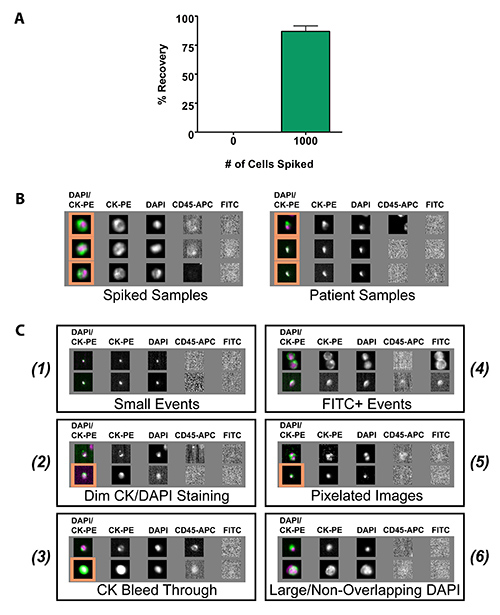
The sensitivity and specificity of the CSS has been well documented in the literature. However, to validate equivalent CTC recovery, spiked (1,000 LNCaP human prostate cancer cells) and unspiked human blood samples from healthy volunteer donors were processed on the CSS using the standard CSSCTC protocol. As expected, unspiked samples were free of CTCs, 0.00±0.00%, and CTC recovery was demonstrated to be 86.9±4.71% for spiked samples (Figure 1A). CSS gallery images obtained from spiked samples were of optimal quality and CTCs were easy to distinguish from non-CTCs. However, when processing samples obtained from cancer patients, identification of CTCs is slightly more challenging, with many cells appearing smaller in size and being less easily distinguishable from non-CTCs (Figure 1B). In addition, when reviewing patient samples 6 categories of events were identified that were commonly discrepant items between reviewers (Figure 1C). These 6 categories included, (1) small events that did not meet the 4 µm size requirement for CTC classification; (2) items with dim CK and/or DAPI staining; (3) justified (should be counted as a CTC) versus unjustified (should not be counted as a CTC) bleed through into the CD45-APC channel caused by bright CK-PE staining; (4) FITC+ events; (5) pixelated images in the CK and/or DAPI channels; and (6) events with DAPI staining that is larger than CK images or those with DAPI staining that does not overlap >50% with the CK image. For categories (2) and (5) specific criteria exist for CTC classification. For category (2), items with dim CK/DAPI can be classified as CTCs provided that an intact membrane can be observed in the CK channel and an appropriately sized DAPI image is noted. For category (5), items with pixelated CK/DAPI cannot be classified as CTCs if any pixelation is observed in the CK channel. However, pixelation is acceptable in the DAPI channel provided that it is not too severe (i.e. image is entirely white on a background, no grey areas - described by Janssen Diagnostics (formerly Veridex) as white paint on a black background) or diffuse (must still appear oval in shape and fit within the CK).
User-Defined Marker Assay Development
Adaptation of the CSS to characterize CTCs for user-defined markers requires significant work-up with rigorous controls and has been described previously16. As a general rule, appropriate optimization of any user-defined marker requires that negative controls be employed to ensure that results are specific. The best results are obtained when spiked samples are processed with both a nonspecific IgG control in place of the target antibody and with the antibody diluent alone as described previously. Various target antibody concentrations and exposure times should also be assessed and validated using cell lines with high, low, and absent (negative) antigen densities. Optimal protocol conditions are achieved when the assay demonstrates both high sensitivity for the target antigen and low background noise from nonspecific binding16.
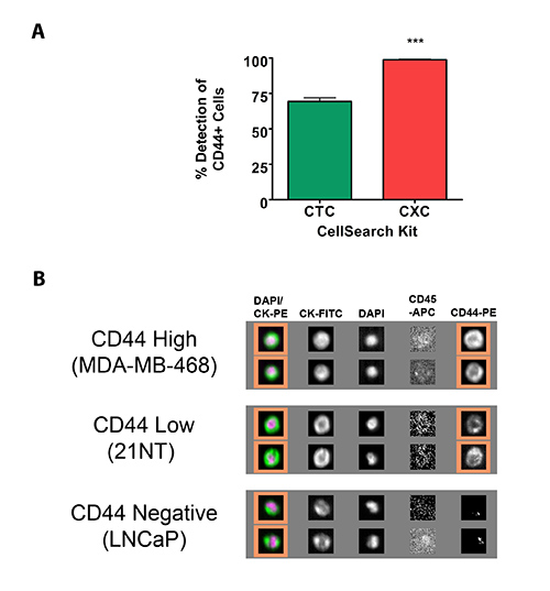
An example of this work-up using a cancer stem cell marker, CD44, is presented here. Initial testing with this marker began using the standard CSS CTC kit (hereafter referred to as the traditional CTC kit), which utilizes the FITC channel for user-defined marker development. Using the traditional CTC kit, it was demonstrated that, after significant optimization, the maximum number of CTCs that could be classified as CD44+ was 69.3±2.67% using samples spiked with 1,000 MDA-MB-468 human breast cancer cells, known to demonstrate high CD44 expression with the majority of cells (98.4±0.90%; as determined by flow cytometry) expressing this protein (Figure 2A). Based upon these findings it was hypothesized that the commercially available CSS CXC kit might produce improved results. This kit allows for improved visualization of markers with a lower antigen density (~50,000 antigens/cell) compared with the traditional CTC kit (optimized for markers with a density of ~100,000 antigens/cell) by reversing the fluorescence channel in which the CK8/18/19 (traditionally represented in the PE channel) and the user's marker of interest (traditionally represented in the FITC channel) are represented (therefore hereafter the CXC kit will be referred to as the low antigen density CTC kit)26. After significant optimization, it was demonstrated that this change allowed for improved CD44 staining, with 98.8±0.51% of CTCs classified as CD44+ using CD44-PE at a concentration of 1.0 µg/ml and an exposure time of 0.6 sec (Figure 2A). Appropriate optimization of any user-defined marker also requires validation using high antigen density (MDA-MB-468), low antigen density (21 NT), and negative (LNCaPs) cell lines for the marker of interest (Figure 2B).
CTC Analysis in Preclinical Mouse Models
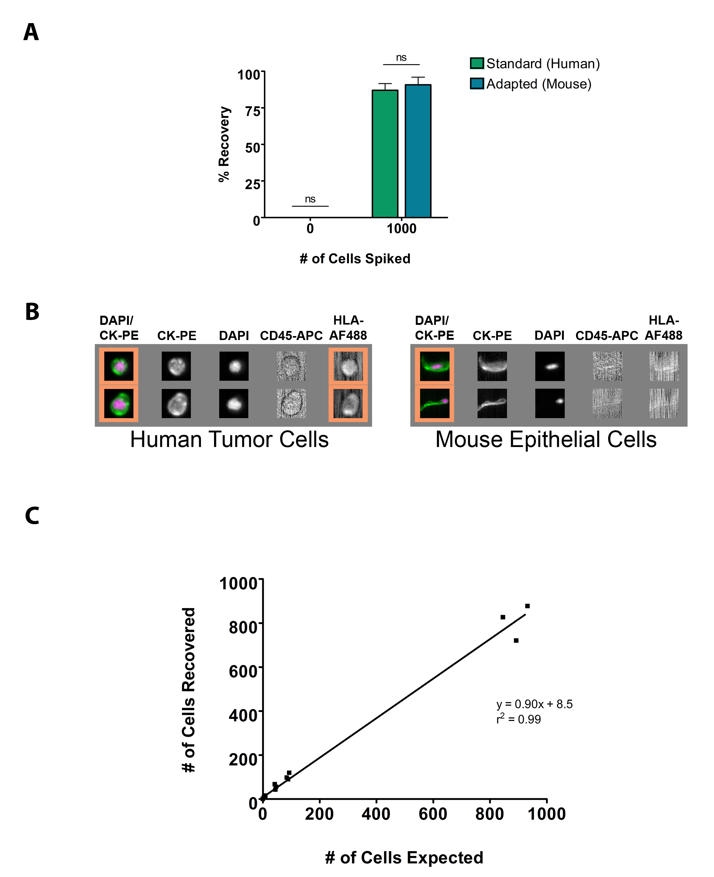
To determine the sensitivity and specificity of the adapted mouse CSS protocol, spiked (1,000 LNCaP human prostate cancer cells) and unspiked mouse blood samples were processed manually and scanned on the analysis instrument and compared to results obtained using the same cell line processed using the standard automated CSS protocol on the preparation instrument (Figure 3A). As expected, unspiked samples were free of CTCs using both assays, 0.00±0.00% and CTC recovery using the adapted mouse kit (90.8±5.18%) was not significantly different from results obtained using the standard automated system (86.9±4.71%; p > 0.05). Images obtained using the manual mouse adapted protocol did not differ from those observed using the standard automated technique, with the exception of the addition of the HLA-FITC marker. In addition, mouse squamous epithelial cells do not stain positively for HLA-FITC (Figure 3B). To confirm that this technique was as sensitive as the standard CSS protocol for the isolation of low number of CTCs, serial dilutions were performed with spiked blood samples and the correlation of expected number of cells versus recovered number of cells was assessed (Figure 3C). Results demonstrate that CTCs could effectively be recovered down to a sensitivity of 5 cells/50 µl of whole blood using this assay. These values correlated with expected results with an r2 = 0.99.

Figure 1. CTC enumeration and interpretation using the standard CSS protocol. (A) CTC recovery measured as a percentage of the number of spiked cells. Cells were counted by hemocytometer and ~1,000 LNCaP human prostate cancer cells were spiked into 7.5 ml of human blood. Unspiked human blood samples were used as a negative control (n = 3). Data are presented as the mean ± SEM. (B) Representative CSS gallery images of the differences in CTC quality observed in spiked blood samples (i.e. healthy donor blood spiked with tumor cells from culture) versus samples collected from cancer patients. (C) Representative CSS gallery images of commonly discrepant items that are often misclassified. Orange squares indicate acceptable CTCs, identified as CK+/DAPI+/CD45-. Images acquired at 10X objective magnification. Click here to view larger image.

Figure 2. Characterization of CTCs for user-defined markers using the CSS. (A) Percentage of cells classified as CD44+ using the CTC and CXC kits on the CSS (n = 3). Data are presented as the mean ± SEM. *** = significantly different (P < 0.0005). (B) Representative CSS gallery images of blood from a healthy volunteer donor (7.5 ml), spiked with ~1,000 cells from the identified cell line, incubated with 1.5 µg/ml of anti-CD44-PE, and scanned at an exposure time of 0.2 sec. Orange squares indicate CD44+ CTCs, identified as CK+/DAPI+/CD45-/CD44-PE+. Click here to view larger image.

Figure 3. Adaptation of the CSS procedure for use in preclinical mouse models of metastasis. (A) CTC recovery using the adapted mouse CSS protocol measured as a percentage of the number of spiked cells and compared to results obtained using the standard human CSS protocol. Cells were counted by hemocytometer and ~1,000 LNCaP human prostate cancer cells were spiked into 7.5 ml of human blood. Unspiked human blood samples were used as a negative control (n = 3). Data are presented as the mean ± SEM. ns = not significant. (B) Representative CSS gallery images of CTCs captured the adapted mouse CSS protocol demonstrating that HLA-AF488 is able to distinguish human from mouse cells. (C) Analysis of correlation and linear regression of the expected versus recovered number of spiked tumor cells at various concentrations. LNCaP human prostate cancer cells were initially counted by hemocytometer and subsequently spiked into mouse blood at a concentration of ~1,000 tumor cells/50 µl of blood. Spiked mouse blood was then serially diluted to a concentration of 5 tumor cells/50 µl and processed using the mouse adapted CSS protocol (n = 3). Click here to view larger image.
| # of Samples with User Defined Marker Added | Total Volume to Add to Reagent Cup (µl) |
| 1 | 450 |
| 2 | 600 |
| 3 | 750 |
| 4 | 900 |
| 5 | 1,050 |
| 6 | 1,200 |
| 7 | 1,350 |
| 8 | 1,500 |
Table 1. Total volume requirements for the CSS when processing various numbers of samples with a user-defined marker. **Adapted from Veridex White Paper (available at: http://www.veridex.com/pdf/CXC_Application_Guideline.PDF).
Dyskusje
Despite the development of many new CTC technologies since the introduction of the CSS in 2004, this technique is still the only clinically approved technology on the market today and therefore it is considered the current gold standard for CTC detection and enumeration. This manuscript has demonstrated that although the CSS has rigorous quality control standards it can be subject to interpretation bias and that CTC identification in patient samples is much different from identification in spiked samples. Six categories of commonly discrepant items were identified that can cause CTC misclassifications to occur. These discrepant items highlight the need for multiple reviewers on each patient sample processed on this instrument. In addition, the differences observed in spiked versus patient obtained CTCs demonstrates that there is a necessity for any new CTC technologies to be validated in both spiked and patient samples. In addition, these new technologies must be compared to the gold standard CSS using split sample testing of both spiked and patient samples, as efficient CTC capture from spiked samples only does not necessarily reflect CTC capture efficiency in patient samples.
Although the CSS has the capability to perform characterization of captured CTCs, it is quite restricted with regards to highly customizable optimization. In general, the only parameters that can be changed on this instrument for optimization of user-defined markers are the antibody concentration and the length of time that the fluorophore is exposed to the mercury lamp. This limited capacity for optimization can present problems when working-up user-defined markers on the CSS. One solution proposed in this manuscript (described in detail previously16) is the use of the low antigen density CTC kit which switches the FITC and PE fluorescent channels allowing for better visualization of markers with a low antigen density. Regardless of which kit is utilized (traditional- or low antigen density CTC kit) there are several necessary steps that must be undertaken to ensure appropriate marker sensitivity, specificity, and optimization. Firstly, assay sensitivity must be assessed in comparison to a well validated method, such as flow cytometry, that will allow determination of the expected detection level (i.e. the percentage of cells in the cell population that express the marker of interest) of the user-defined marker. Secondly, the assay must be assessed for its ability to detect the marker of interest in cell lines with various levels of expression (i.e. high and low antigen densities) and its specificity must be validated in a cell line that is negative for the marker of interest. In all cases, all cell lines must be tested using a cells only control (no antibody added), the appropriate IgG control, and the antibody of interest at various concentrations and exposure times to determine the most appropriate settings that will ensure optimization of the user-defined marker. However, it should be noted that although characterization of CTCs is possible on the CSS, currently only one user-defined marker of interest can be explored in each sample, and that the system is very limited with regards to downstream applications due to the harsh processing of the samples.
The unique bedside-to-benchtop approach utilized in CTC research has allowed for quick entry of this useful assay into the clinical setting. However, it has resulted in an inadequate understanding of the basic biology of these rare cells. Therefore the development and optimization of assays that allow for assessment of CTCs in preclinical in vivo mouse models of cancer are needed. This manuscript describes an adapted CSS protocol that allows for CTCs to be assessed in 50 µl volumes of mouse blood, ideally suited for serial CTC collection experimental models. This manuscript demonstrates that CTC enumeration using this protocol is comparable to results obtained using the CSS in combination with the traditional CTC kit, with no significant differences in CTC capture efficiency between the automated and manual separation techniques. In addition, during the development of this assay it was recognized, as previously described in the literature25, that mouse squamous epithelial cell contamination can make accurate identification of CTCs more difficult and sometimes impossible. Therefore to combat this issue a user-defined HLA-Alexa Fluor 488 was added to this protocol to ensure that only human cells (CK+DAPI+CD45-HLA-Alexa Fluor 488+) are being appropriately assigned as CTCs. It is important to note that the LNCaP cell line used in this manuscript is HLAlow and therefore HLA-Alexa Fluor 488 may need to be titrated for cell lines with varying HLA expression. Although we have added an HLA-Alexa Fluor 488 to our protocol to ensure accurate identification of CTCs, it is noteworthy that the vast majority of mouse squamous epithelial cells were easily identifiable by morphology and were limited in cell number (0.33±0.24 events/50 µl; n = 9). Higher cell numbers (up to 11 events/50 µl) were only observed when blood collection (via cardiac puncture) proved difficult and repeated attempts were necessary. Therefore we propose that if desired, on-system characterization of CTCs could be accomplished by omitting this marker. In addition, although not described here, we anticipate that collection of CTCs and further downstream characterization of CTCs using other methods could be easily achieved as demonstrated previously using the CSS27,28.
Although the CSS has been utilized clinically to effectively detect CTCs in the blood of metastatic breast, prostate and colorectal cancer patients4,10,29, it does have several limitations. In up to 35% of patients with various metastatic cancers, CTCs are undetectable despite the presence of widespread systemic disease30. This lack of detection has been proposed to be as a result of the epithelial-to-mesenchymal transition, a well-documented process known to enhance cancer invasion, metastasis, and overall aggressiveness31. This transition has been associated with a significant reduction in epithelial markers, such as EpCAM, and a corresponding increase in mesenchymal markers32. Several studies have recently demonstrated that the presence of these mesenchymal markers in CTCs are indicative of poorer prognosis and that many of these cells lack expression of epithelial markers that would be necessary for their detection using the CSS24,33-38. This suggests that the standard CSS definition may be missing some of the most aggressive CTCs.
Despite the described limitations, it is anticipated the protocols described in this manuscript will be important tools for improved CTC analysis using the CSS, development of novel CTC technologies, optimization of user-defined markers, and improved understanding of CTC biology using in vivo preclinical mouse models. Taken together, these protocols will provide a useful resource for users of this platform interested in preclinical and clinical research pertaining to metastasis and CTCs.
Ujawnienia
The author (A.L.A.) received funding that was provided by Janssen Oncology, whose parent company Johnson & Johnson also owns Janssen Diagnostics LLC, which produces reagents and instruments used in this article.
Podziękowania
This work was supported by grants from the Ontario Institute of Cancer Research (#08NOV230), the Canada Foundation for Innovation (#13199), Prostate Cancer Canada, Janssen Oncology, the London Regional Cancer Program, and donor support from John and Donna Bristol through the London Health Sciences Foundation (to A.L.A.). L.E.L. is supported by a Frederick Banting and Charles Best Canada Graduate Scholarship Doctoral Award. A.L.A is supported by a CIHR New Investigator Award and an Early Researcher Award from the Ontario Ministry of Research and Innovation.
Materiały
| Name | Company | Catalog Number | Comments |
| 0.5 M EDTA | |||
| Anti-human CD44-FITC | BD Pharmigen | 555478 | |
| Anti-human CD44-PE | BD Pharmigen | 555479 | |
| Anti-human HLA-Alexa Fluor 488 | BioLegend | 311415 | |
| Anti-mouse CD45-APC | eBioscience | 17-0451-82 | |
| Bond Primary Antibody Diluent | Leica | AR9352 | |
| CellSave Preservative Tubes | Veridex | 952820 (20 pack) 79100005 (100 pack) | |
| CellSearch CTC Control Kit | Veridex | 7900003 | |
| CellSearch CTC Kit | Veridex | 7900001 | |
| CellSearch CXC Control Kit | Veridex | 7900018RUO | |
| CellSearch CXC Kit | Veridex | 7900017RUO | |
| Instrument Buffer | Veridex | 7901003 | |
| Streck Cell Preservative (aka CytoChex) | Streck | 213350 | |
| 1 ml Syringe | |||
| 10 ml Serological pipette | |||
| 1,000 µl Pipette | |||
| 1,000 µl Pipette tips | |||
| 12 mm x 75 mm Flow tubes | |||
| 200 µl Gel loading tips | |||
| 200 µl Pipette | |||
| 22 G Needle | |||
| 5 3/4 in Disposible Pasteur pipet | VWR | 14672-200 | |
| 5 ml Serological pipette | |||
| Automated pipettor | |||
| Capillary Blood Collection Tube (EDTA) | BD Microtainer | 365974 | |
| CellSearch Analyzer II | Veridex | 9555 | Includes magnests and verification cartirdges |
| CellSearch AutoPrep System | Veridex | 9541 | |
| Centrifuge | |||
| MagCellect Magnet | R&D Systems | MAG997 | |
| Small Latex Bulb | VWR | 82024-550 | |
| Vortex |
Odniesienia
- Siegel, R., Naishadham, D., Jemal, A. Cancer statistics. 63 (1), 11-30 (2013).
- Chambers, A. F., Groom, A. C., MacDonald, I. C. Dissemination and growth of cancer cells in metastatic sites. Nat. Rev. Cancer. 2 (8), 563-572 (2002).
- Pantel, K., Brakenhoff, R. H. Dissecting the metastatic cascade. Nat. Rev. Cancer. 4 (6), 448-456 (2004).
- Cristofanilli, M., Budd, G. T., et al. Circulating tumor cells, disease progression, and survival in metastatic breast cancer. New Engl. J. Med. 351 (8), 781-791 (2004).
- De Bono, J. S., Scher, H. I., et al. Circulating tumor cells predict survival benefit from treatment in metastatic castration-resistant prostate cancer. Clin. Cancer Res. 14 (19), 6302-6309 (2008).
- Cohen, S. J., Ja Punt, C., et al. Prognostic significance of circulating tumor cells in patients with metastatic colorectal cancer. Ann. Oncol. 20 (7), 1223-1229 (2009).
- Hayes, D. F., Cristofanilli, M., et al. Circulating tumor cells at each follow-up time point during therapy of metastatic breast cancer patients predict progression-free and overall survival. Clin. Cancer Res. 12 (14), 4218-4224 (2006).
- Budd, G. T., Cristofanilli, M., et al. Circulating tumor cells versus imaging--predicting overall survival in metastatic breast cancer. Clin. Cancer Res. 12 (21), 6403-6409 (2006).
- Olmos, D., Arkenau, H. -. T., et al. Circulating tumour cell (CTC) counts as intermediate end points in castration-resistant prostate cancer (CRPC): a single-centre experience. Ann. Oncol. 20 (1), 27-33 (2009).
- De Bono, J. S., Scher, H. I., et al. Circulating tumor cells predict survival benefit from treatment in metastatic castration-resistant prostate cancer. Clin. Cancer Res. 14 (19), 6302-6309 (2008).
- Allan, A. L., Keeney, M. Circulating tumor cell analysis: technical and statistical considerations for application to the clinic. J. Oncol. 2010, 426218 (2010).
- Lowes, L. E., Goodale, D., Keeney, M., Allan, A. L. Image cytometry analysis of circulating tumor cells. Methods Cell Biol. 102, 261-290 (2011).
- Lianidou, E. S., Markou, A. Circulating tumor cells in breast cancer: detection systems, molecular characterization, and future challenges. Clin. Chem. 57 (9), 1242-1255 (2011).
- Alix-Panabières, C., Pantel, K. Circulating tumor cells: liquid biopsy of cancer. Clin. Chem. 59 (1), 110-118 (2013).
- Parkinson, D. R., Dracopoli, N., et al. Considerations in the development of circulating tumor cell technology for clinical use. J. Transl. Med. 10, 138 (2012).
- Lowes, L. E., Hedley, B. D., Keeney, M., Allan, A. L. User-defined protein marker assay development for characterization of circulating tumor cells using the CellSearch system. Cytomet. A. 81 (11), 983-995 (2012).
- Fehm, T., Müller, V., et al. HER2 status of circulating tumor cells in patients with metastatic breast cancer: a prospective, multicenter trial. Breast Cancer Res. Treat. 124 (2), 403-412 (2010).
- Liu, Y., Liu, Q., et al. Circulating tumor cells in HER2-positive metastatic breast cancer patients: a valuable prognostic and predictive biomarker. BMC Cancer. 13, 202 (2013).
- Slamon, D. J., Leyland-Jones, B., et al. Use of chemotherapy plus a monoclonal antibody against HER2 for metastatic breast cancer that overexpresses HER2. New Engl. J. Med. 344 (11), 783-792 (2001).
- Marty, M., Cognetti, F., et al. Randomized phase II trial of the efficacy and safety of trastuzumab combined with docetaxel in patients with human epidermal growth factor receptor 2-positive metastatic breast cancer administered as first-line treatment: the M77001 study group. J. Clin. Oncol. 23 (19), 4265-4274 (2005).
- Slamon, D., Eiermann, W., et al. Adjuvant trastuzumab in HER2-positive breast cancer. New Engl. J. Med. 365 (14), 1273-1283 (2011).
- Welch, D. R. Technical considerations for studying cancer metastasis in vivo. Clin. Exp. Metast. 15 (3), 272-306 (1997).
- Goodale, D., Phay, C., Postenka, C. O., Keeney, M., Allan, A. L. Characterization of tumor cell dissemination patterns in preclinical models of cancer metastasis using flow cytometry and laser scanning cytometry. Cytometry A. 75 (4), 344-355 (2009).
- Gorges, T. M., Tinhofer, I., et al. Circulating tumour cells escape from EpCAM-based detection due to epithelial-to-mesenchymal transition. BMC Cancer. 12, 178 (2012).
- Eliane, J. -. P., Repollet, M., et al. Monitoring serial changes in circulating human breast cancer cells in murine xenograft models. Cancer Res. 68 (14), 5529-5532 (2008).
- Flores, L. M., Kindelberger, D. W., et al. Improving the yield of circulating tumour cells facilitates molecular characterisation and recognition of discordant HER2 amplification in breast. 102 (10), 1495-1502 (2010).
- Sieuwerts, A. M., Kraan, J., et al. Molecular characterization of circulating tumor cells in large quantities of contaminating leukocytes by a multiplex real-time PCR. Breast Cancer Res. Treat. 118 (3), 455-468 (2009).
- Cohen, S. J., Ja Punt, C., et al. Relationship of circulating tumor cells to tumor response, progression-free survival, and overall survival in patients with metastatic colorectal cancer. J. Clin. Oncol. 26 (19), 3213-3221 (2008).
- Allard, W. J., Matera, J., et al. Tumor cells circulate in the peripheral blood of all major carcinomas but not in healthy subjects or patients with nonmalignant diseases. Clin. Cancer Res. 10 (20), 6897-6904 (2004).
- Thiery, J. P., Acloque, H., Huang, R. Y. J., Nieto, M. A. Epithelial-mesenchymal transitions in development and disease. Cell. 139 (5), 871-890 (2009).
- Yang, J., Weinberg, R. A Epithelial-mesenchymal transition: at the crossroads of development and tumor metastasis. Dev. Cell. 14 (6), 818-829 (2008).
- Gradilone, A., Raimondi, C., et al. Circulating tumour cells lacking cytokeratin in breast cancer: the importance of being mesenchymal. J. Cell. Mol. Med. 15 (5), 1066-1070 (2011).
- Königsberg, R., Obermayr, E., et al. Detection of EpCAM positive and negative circulating tumor cells in metastatic breast cancer patients. Acta Oncol. 50 (5), 700-710 (2011).
- Kasimir-Bauer, S., Hoffmann, O., Wallwiener, D., Kimmig, R., Fehm, T. Expression of stem cell and epithelial-mesenchymal transition markers in primary breast cancer patients with circulating tumor cells. Breast Cancer Res. 14 (1), (2012).
- Mego, M., Mani, S. A., et al. Expression of epithelial-mesenchymal transition-inducing transcription factors in primary breast cancer: The effect of neoadjuvant therapy. Int. J. Cancer. 130 (4), 808-816 (2012).
- Yu, M., Bardia, A., et al. Circulating breast tumor cells exhibit dynamic changes in epithelial and mesenchymal composition. Science. 339 (6119), 580-584 (2013).
- Armstrong, A. J., Marengo, M. S., et al. Circulating tumor cells from patients with advanced prostate and breast cancer display both epithelial and mesenchymal markers. Mol. Cancer Res. 9 (8), 997-1007 (2011).
Przedruki i uprawnienia
Zapytaj o uprawnienia na użycie tekstu lub obrazów z tego artykułu JoVE
Zapytaj o uprawnieniaThis article has been published
Video Coming Soon
Copyright © 2025 MyJoVE Corporation. Wszelkie prawa zastrzeżone