Method Article
Combination of High Ligation and Intraoperative Embolization using Polidocanol for Treatment of Varicoceles
In This Article
Summary
To reduce the postoperative recurrence rate of varicocelectomy, we combined high ligation of varicocele with intraoperative embolization. We injected polidocanol from the spermatic vein during surgery to embolize the branches of the spermatic vein and collateral veins. This is an alternative surgical method for the treatment of varicocele.
Abstract
Varicoceles are dilated veins within the pampiniform plexus and are relatively common in the general male population. The spermatic vein has many branches in the scrotal segment and then gradually merges into 1-2 trunks after passing through the internal inguinal ring. The key to a successful varicocelectomy is to ligate all the spermatic veins while protecting the testicular arteries and spermatic lymphatic vessels from damage. The small veins, including the branches of spermatic veins and collateral veins, are easily missed for ligation during conventional high ligation of varicocele, which has been suggested as a major cause of postoperative recurrence. Although microsurgery effectively reduces the risk of missing ligation of the spermatic veins during surgery, it has several shortcomings, such as long operation time and a steep learning curve. More importantly, this technique is difficult to carry out in primary hospitals due to the requirement of specialized equipment. Therefore, an attempt to modify the traditional high ligation aiming to reduce the postoperative recurrence rate has been carried out here. The protocol here combines traditional high ligation with intraoperative embolization to seal off the branches of the spermatic vein and collateral veins. We rapidly injected foamed sclerosant into the internal spermatic vein under direct observation after separation of the spermatic vein and then ligated all the veins. The foamed sclerosant through the varicose vein hampers endothelial cell growth, promotes the growth of thrombus and fibrosis, and ultimately forms fibrous streaks that permanently fill the venous. The results showed a more satisfactory effect on reducing the postoperative recurrence rate compared with traditional high ligation. Since this protocol is simple to carry out and has better results in reducing the recurrence rate, this can be an alternative surgical method for the treatment of varicocele, especially in primary hospitals.
Introduction
Varicoceles, with an incidence of approximately 15%-20% in the general male population and 35%-40% among the infertile population, is one of the major causes of infertility1,2,3. In addition, varicoceles can cause pain and discomfort and a decline in androgen levels4. In recent decades, different surgical procedures have been consistently applied to treat varicoceles, including high ligation of varicocele, inguinal and sublingual micro varicocelectomy, laparoscopic spermatic vein ligation, and interventional embolization5. High retroperitoneal ligation of the spermatic vein is the traditional surgical procedure used to treat varicoceles6. Moreover, it is the simplest surgical procedure for the surgical treatment of varicocele and is easy to carry out at any center. However, this method easily misses ligating the branch veins, which can subsequently lead to postoperative recurrence. Laparoscopic ligation of the spermatic vein may risk damaging the abdominal organs. Furthermore, the risk of arterial damage and the requirement for specialized equipment are high. It has been demonstrated that micro varicocelectomy produces better results than other surgical procedures in improving semen quality and reducing the postoperative recurrence rate. Therefore, micro varicocelectomy has been considered as the golden standard of surgical treatments for varicocele. However, this technique has several shortcomings, such as long operation time and a steep learning curve. More importantly, the requirement for specialized equipment and the difficulty in carrying out the technique in primary hospitals.
We present a new surgical procedure that is not limited by the equipment and, thus, can be applied in any center. We also expect this procedure to achieve a better effect on reducing the postoperative recurrence rate. Based on the procedure of traditional high ligation, we applied intraoperative embolization simultaneously with high ligation. We injected embolic agents from the internal spermatic vein under direct observation during surgery to embolize the branches of spermatic vein, aiming to cover the possibility of missing ligation. The procedure may block the veins as completely as possible and reduce the postoperative recurrence rate. Since the procedure derives from traditional high ligation of spermatic vein, there is no limit of equipment, and it is easy to be performed by any surgeon and can be carried out in most centers.
High ligation combined with intraoperative embolization (HLIE) has a short learning curve and can lead to complete occlusion of the spermatic vein. After being reviewed and approved by the First Affiliated Hospital, Sun Yat-sen University, on January 10, 2013, we began applying HLIE and evaluating its outcomes in comparison with traditional high ligation.
Protocol
All procedures in the following protocol were reviewed and approved by the First Affiliated Hospital, Sun Yat-sen University.
1. Patient preparation
- Apply the following inclusion criteria: male sex; varicoceles during physical examination and on ultrasonography; infertility or abnormal semen or unrelieved pain by drugs.
- Apply the following exclusion criteria: other known causes of male infertility, such as cryptorchidism, cancer, scrotum and reproductive tract surgery, hypogonadism, and azoospermia.
- Perform a testicle and spermatic cord palpation and ultrasound examination to determine the classification of varicocele. Place the patient in a standing position and confirm whether there is a dilated and clumped venous plexus on the scrotum skin.
- Examine the patient's spermatic cord to confirm whether the spermatic veins dilate during spontaneous respiration and Valsalva's movements (after deep inhalation, close the glottis tightly, then exhale forcefully to increase abdominal pressure). Perform an ultrasound examination to evaluate the diameter of the spermatic vein.
- Inform the patient of the benefits and risks of surgery, including excessive bleeding and blood transfusion, testicular atrophy, recurrence, scrotal edema, hydrocele, and postoperative infection, then obtain written consent.
- Carry out the surgery under epidural anesthesia or general anesthesia with the patient placed in a supine position.
2. Pre-HLIE preparation
- Monitor the patient's vital signs carefully by an anesthesiologist during surgery.
- Sterilize the skin of the thigh, perineum, and abdomen 2x with iodophor. Create a longitudinal incision approximately 2-3 cm in length with a scalpel. Establish the incision by standard technique: 2 cm above the internal inguinal ring.
3. High ligation and intraoperative embolization (HLIE)
- After incision of the subcutaneous tissue, cut the aponeurosis of the obliquus externus abdominis approximately 3 cm, and bluntly separate the muscles and transverse fascia below to expose the peritoneum and retroperitoneal adipose tissue.
- Use a retractor to pull the muscles and peritoneum aside to have a clear view. The spermatic cord is just behind the peritoneum, between the peritoneum and retroperitoneal adipose tissue. Use a retractor to pull the adipose tissue aside and find the spermatic cord adjacent to the peritoneum, then bluntly separate the spermatic cord from surrounding connective tissue with forceps.
- Grasp the ipsilateral scrotum by hand and pull the testicle to confirm the spermatic vein. Squeeze the testicle to promote venous drainage, which will be beneficial for intraoperative embolization with polidocanol. Cut the spermatic fascia with scissors to expose the spermatic vein, then bluntly separate the spermatic vein with forceps.
NOTE: During surgical dissection of the spermatic vein, any injury to the accompanying arteries and lymphatic vessels should be avoided. - Prepare vascular hardening agent for embolization: Use two 10 mL sterile syringes to mix 5 mL of polidocanol and 5 mL of air. Do this until a 10 mL polidocanol foam sclerosant mixture is obtained; use this for embolization.
- Make an approximately 0.2-0.3 cm longitudinal incision with a scalpel on the spermatic vein. Insert a 0.8 cm sterile syringe needle into the spermatic vein.
- Inject the 10 mL polidocanol foam sclerosant mixture rapidly into the distal segment of the spermatic vein through the needle.
- Remove the needle, and double ligate the spermatic vein with 3-0 non-absorbable suture line at sites approximately 0.5 cm proximal and distal to the incision of the vein.
- Finally, check whether there is any bleeding at any point, including the spermatic cord, retroperitoneal adipose tissue, and muscles. If yes, stop the bleeding with an electrocoagulation electrotome carefully. Suture the abdominal incision with 3-0 absorbable suture material.
4. Postoperative management
- Document the intra- and post-operative parameters carefully, including the diameters of the spermatic vein according to the results of ultrasound examination, operation time (from the beginning of incision to the end of suturing), and blood loss (calculated by vacuum drainage).
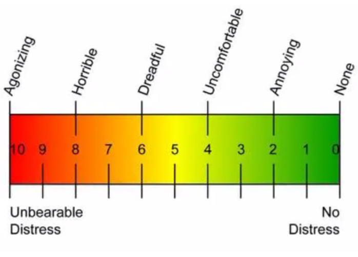
- Perform patient follow-up after surgery at 1 month, 3 months, 6 months, and 1 year. Perform testicle examinations and ultrasound, pain scores (the visual analog scale (VAS) pain score to evaluate the pain of varicocele patients before and after surgery; Figure 1), semen analysis, and whether the patients had achieved impregnation or experienced scrotal edema, varicocele recurrence, or testicular atrophy.
- Asses the effect of treatment, including semen parameters and pain score, recurrence rate, and probability of complications of HLIE, and perform statistical analysis with SPSS software, taking p < 0.05 as statistically significant.
Results
HLIE (Figure 2) was performed on 53 patients, and traditional HL was performed on 81 patients from 2013 to 2019. The mean ages of the two groups were 31.29 years (range 15 to 65) and 29 years (range 15 to 64), respectively. A total of 79.1% (n=106) of the patients were treated for a left-sided varicocele in this sample. The most common clinical grades are Grade II and Grade III (Grade II: venous clusters cannot be seen on the surface of the scrotum, but the spermatic vein is varicose by palpation; Grade III: venous clusters can be seen on the surface of the scrotum; Table 1).
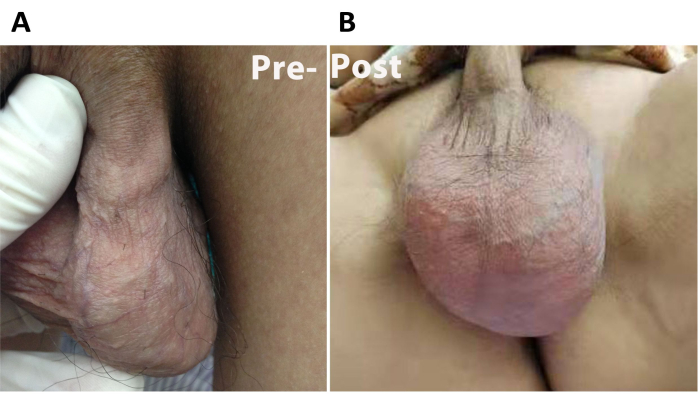
Data from 9 patients are presented here. There were 2 patients in the HLIE group and 7 patients in the HL group having clinical recurrence during follow-up. The patients treated with HLIE had a postoperative recurrence rate of 3.77% and an incidence of scrotal edema of 18.87%. The recurrence rate and incidence of scrotal edema in patients treated with HL were 8.64% and 7.41%, respectively. The recurrence rate of HLIE was lower than that of HL; however, we did not find a significant difference in the recurrence rate between the two groups, which may be related to the sample size. The overall success rates of pain-relieving were 82.35% in the HLIE group and 68.75% in the HL group, with a median follow-up of 18 months. Most of the patients' pain symptoms were significantly improved after the operation, which demonstrated that HLIE had a satisfactory effect on relieving the pain of varicocele. A total of 53 patients underwent the HLIE procedure, and none of the patients had venous clusters on their scrotum surface during physical examination 1 month after surgery (Figure 3). The only complication was scrotal edema, which resolved spontaneously within 1 month after surgery. No other severe complications, such as testicular atrophy, occurred during follow-up.

Figure 1: VAS score. The visual analog scale (VAS) pain score is used to evaluate the pain of varicocele patients before and after surgery. Please click here to view a larger version of this figure.

Figure 2: Protocol for HLIE. (A) Incision of the subcutaneous tissue. (B) Locate the spermatic vein and insert a syringe needle. (C) Use two syringes to mix polidocanol and air. (D) Rapidly inject polidocanol foam sclerosant mixture into the spermatic vein. Please click here to view a larger version of this figure.

Figure 3: Images of the patient's scrotum before and after the operation. (A) Patient's scrotum before the operation, venous clusters can be seen on the surface of the scrotum. (B) Patient's scrotum 1 month after operation. Disappearance of the venous masses on the surface of the scrotum. Please click here to view a larger version of this figure.
| Patients | HLIE group (n=53) | HL group (n=81) | P |
| age(year) | 31.29±10.05 | 29±8.499 | 0.0937 |
| recurrence | 2/53(3.77%) | 7/81(8.64%) | 0.4822 |
| Pain relieving | 28/34(82.35%) | 33/48(68.75%) | 0.2043 |
| Scrotal edema | 10/53(18.87%) | 6/81(7.41%) | 0.0579 |
Table 1: Comparison of the outcomes between groups. There were 2 patients in the HLIE group and 7 in the HL group having clinically significant recurrence during follow-up. The results of the pre-and post-operative tests are shown here.
Discussion
This protocol presents a new surgical method for varicocele that combines traditional high ligation with intraoperative embolization to seal off the internal spermatic vein branches sufficiently. We expect to reduce the possibility of missing ligation of the small branches to reduce the postoperative recurrence rate by this method. The follow-up results show that this method can reduce the postoperative recurrence rate and improve the appearance of scrotum covered with varicose veins in the short term. The specific procedures are described as follows. The patient was placed in a supine position. A skin incision was made approximately 2-3 cm above the internal inguinal ring. After the subcutaneous tissue was incised, the aponeurosis of the obliquus externus abdominis was cut, the muscles were separated with a blunt dissection, and the transverse fascia below this area was cut. The muscles and peritoneum were then pulled aside by a retractor to locate the spermatic vein. After the spermatic vein was separated, a 0.8-cm sterile syringe needle was inserted approximately 1.5-2.0 cm into the spermatic vein. Two 10 mL sterile syringes were used to mix 5 mL of polidocanol and 5 mL of air. A 10 mL polidocanol foam sclerosant mixture was rapidly injected into the lower segment of the spermatic vein through the needle inserted into the spermatic vein. Then, the sites approximately 0.5 cm proximal and distal to the incision of the vein were double-ligated. After the bleeding was carefully stopped, the abdominal incision was sutured. In our protocol, the initial steps are roughly the same as those of traditional high ligation. The difference is that we inject the vascular hardening agent into the internal spermatic vein during the operation to embolize the internal spermatic vein branches and collateral veins, which may be missing ligated during surgery and cause postoperative recurrence. After the improvement, the overall steps are relatively simple, efficient, and easy to popularize. More importantly, no limitations of this technique exist as long as varicocelectomy can be performed.
Since Tulloch et al.7 reported that spermatic vein ligation could improve the semen quality of patients with varicoceles in 1955, many approaches and techniques have been applied to treat varicoceles. The current main surgical procedures for varicocele include Palomo surgery, laparoscopic surgery, interventional embolization (anterograde and retrograde), and microsurgery5,8,9. The key to a successful surgery is to ligate all the spermatic veins while protecting the testicular arteries, spermatic lymphatic vessels, and vas deferens from damage.
Due to its high resolution and facilitation of the preservation of lymphatic vessels and arteries10, microsurgery effectively reduces arterial and lymphatic damage while simultaneously reducing the risk of missing ligation of the spermatic vein during surgery. In recent years, many clinical studies, as well as systematic reviews and meta-analyses, have also shown that micro varicocelectomy produced better results than high retroperitoneal ligation for improving semen quality, increasing natural conception rates, and reducing the complication and recurrence rates of varicoceles11,12,13,14 . However, microsurgical techniques have several shortcomings, such as a long operation time and a steep learning curve15. More importantly, the requirement for equipment is extremely high, and this technique is consequently difficult to carry out in primary hospitals. Laparoscopic ligation of the spermatic vein is performed in the abdominal cavity, which risks damaging the abdominal organs. Moreover, the procedure is expensive, the learning curve is long, and the risk of arterial damage is high16, 17.
With the development of interventional radiology, embolization of the internal spermatic vein with an embolus or an injection of sclerosant or foamed sclerosant has become one of the alternative approaches used to treat varicoceles. In addition, these methods cause minor damage, and the postoperative recovery is fast. Many studies have reported that varicocele embolization has beneficial effects on improving semen quality, increasing the pregnancy rate, and relieving pain18,19,20,21. However, embolization using a nickel-titanium screw bolt produces a risk of serious complications caused by embolus. During embolization using sclerosant or foamed sclerosant, because the direction of drug injection is opposite to the blood flow and the pressure of drug injection is low, drugs cannot completely embolize the collateral circulation and venous branches. In addition, this operation should be conducted under X-ray guidance, requires difficult techniques, and can possibly cause radioactive injury to the patient. Therefore, the application of this surgical approach is limited.
The recurrence rate of high ligation of the spermatic vein or embolization is significantly higher than that of microsurgery. Marsman et al. termed varicoceles accompanied by collateral veins aberrantly fed varicoceles (AFVs). Some researchers have suggested that AFVs are a major cause of varicocele recurrence22,23,24. Because collateral veins are connected to the spermatic vein, it is possible to reconstruct the collateral vein if it is not ligated. If back-streaming blood flows to the testicular vein from the lateral branches of the spermatic cord, it can lead to the recurrence of varicoceles. During a second venography, Fenelv et al.23 found that the collateral veins of recurrent patients who had never been embolized or ligated were incompletely blocked.
Therefore, we attempted to treat varicoceles using a new surgical technique, namely, high ligation of the spermatic vein combined with intraoperative embolization with a foamed sclerosant. A high-pressure injection of a foamed sclerosant was used to completely embolize the rami communicans of the spermatic vein and the collateral veins possibly missed during ligation. Injection of sclerosant or foamed sclerosant through the varicose vein may damage endothelial cells in the venous wall25, 26, promote the growth of a thrombus, granulation tissue, and fibrosis in the collapsed vein, and ultimately form fibrous streaks that permanently obstruct the venous lumen, thus achieving the therapeutic purpose by collapsing the varicose veins. After the separation of the spermatic vein during surgery, we rapidly injected foamed sclerosant into the veins with high pressure under direct observation; this approach can effectively eliminate the rami communicans of spermatic and collateral veins, thus reducing the recurrence rate. We attempted to achieve an effect similar to microsurgery in improving semen quality and reducing the recurrence rate of varicoceles after surgery while avoiding some shortcomings of microsurgery, such as the long operation time, steep learning curve, and reduction of the embolus. Additionally, we expect to modify the varicocelectomy procedure for the purpose of making it more efficient and easier to carry out in primary hospitals.
The current reported recurrence rate and incidence of scrotal edema after high ligation surgery are 7.3%-34.9% and 6.4%-10.0%, respectively27,28, and the recurrence rate and incidence of scrotal edema after microsurgery are 0%-3.6% and 0%-2%, respectively14,28,29,30. Cayan et al. compared the recurrence rate and the incidence of scrotal edema of 232 patients treated with high ligation with those of 236 patients treated with microsurgery. Their results showed that patients treated with high ligation had a postoperative recurrence rate of 15.15% and an incidence of scrotal edema of 9.09%. The patients treated with microsurgery had a postoperative recurrence rate of 2.11% and an incidence of scrotal edema of 0.69%, which were significantly lower than those in patients treated with high ligation14. In a study by Ghanem et al.15 of 109 patients who were treated with high ligation, eight had varicocele recurrence, and seven experienced scrotal edema. Among 304 patients treated with microsurgery, none had varicocele recurrence, and five experienced scrotal edema. Therefore, the postoperative recurrence rate and incidence of scrotal edema were significantly lower among patients treated with microsurgery than among those treated with high ligation. With regard to embolization, the study showed that the postoperative recurrence rate was 2-24%15,27,31,32,33,34. A meta-analysis demonstrated that the recurrence rates among patients who were treated with embolization and high ligation were 12.7% and 14.97%, respectively. From 2013 to 2019, we followed 53 patients treated with HLIE and 81 patients treated with HL. The results show that patients treated with HLIE had a postoperative recurrence rate of 3.77% and an incidence of scrotal edema of 18.87%. The recurrence rate and incidence of scrotal edema in patients treated with HL were 8.64% and 7.41%, respectively. The recurrence rate of HLIE was apparently lower than that of HL; however, we did not find a significant difference between the two groups, which may be related to the sample size. HLIE may have advantages over HL in terms of reducing the postoperative recurrence rate.
Due to the surgical ligation of spermatic veins, the varicose vein was not removed, and it required a relatively long time to collapse and regain the normal appearance of the scrotum. Therefore, the surgery did not improve the appearance of the scrotum, which was covered with varicose veins in the short term. It takes weeks or even months to recover the normal appearance of the scrotum, which will bring some psychological burden to the patients. It is possible to improve the appearance of the scrotum by injection of foamed sclerosants, which may adhere to and close spermatic veins in the short term. In the first month after surgery, none of the 53 patients presented varicoceles on their scrotum surface during physical examination, suggesting that this surgical method successfully improves the appearance of the scrotum. However, the postoperative incidence of scrotal edema was higher than the reported incidence for patients treated with high ligation, microsurgery, or embolization. A total of 10 patients exhibited scrotal edema after HLIE, but the edema can spontaneously disappear within 1 month after surgery. During the surgical separation of the spermatic vein, we avoided injuring the accompanying arteries and lymphatic vessels. Therefore, this may be related to the dose of foamed sclerosant injected, or short-term local venous disorders caused by embolization of the rami communicans and collateral circulation. The second European Consensus Meeting on Foam Sclerotherapy recommended that the safe dose of foamed sclerosant was 6-8 mL35. During the surgery, we mixed 5 mL of polidocanol and 5 mL of air evenly to obtain a final volume of 10 mL of foamed sclerosant. Then, we quickly inserted it into the lower segment of the spermatic vein. With adequate pressure, the foamed sclerosant entered the lower venous rami communicans and tiny vein branches. However, because the venous rami communicans were fully embolized, the venous return was obstructed in the short term, leading to a higher incidence of edema. This also verifies the perspective that the occlusion of spermatic veins produced by the surgical procedure is sufficient.
We present this protocol to modify conventional varicocelectomy by combining traditional high ligation with intraoperative embolization, aiming to block the branches of the spermatic vein and collateral veins, which may be a major cause of varicocele recurrence if missing ligated. The results show a satisfactory effect on reducing the postoperative recurrence rate and improving the appearance of the scrotum which was covered with varicose veins. Although the incidence of postoperative edema is higher, edema can spontaneously disappear within 1 month after surgery. Additionally, this procedure is simple and can easily be performed at any center as long as high ligation can be carried out. We expect it to become an alternative surgical method for the treatment of varicocele, especially in primary hospitals.
Disclosures
There are no competing financial interests.
Acknowledgements
This work is funded by the Guangzhou Science and Technology Program-Basic and Applied Basic Research (Grant No. 2023A04J2180).
Materials
| Name | Company | Catalog Number | Comments |
| 3-0 absorbable suture material | Johnson & Johnson-Ethicon | 20203021529 | |
| 3-0 non-absorbable suture line | Johnson & Johnson | 20202020196 | |
| electrocoagulation electrotome | Shinva | 20183010484 | |
| forceps | Shinva | 20140168 | |
| iodophor | ADF Hi-tec Disinfectants Co., Ltd. | 2019029215 | |
| polidocanol | Shanxi Tianyu Pharmaceutical Co., Ltd. | H20080445 | |
| retractor | Shinva | 20150218 | |
| syringes | Medicom | 20153140848 |
References
- Sasson, D. C., Kashanian, J. A. Varicoceles. JAMA. 323 (21), 2210 (2020).
- Templeton, A. Varicocele and infertility. Lancet. 361 (9372), 1838-1839 (2003).
- Kang, C., Punjani, N., Lee, R. K., Li, P. S., Goldstein, M. Effect of varicoceles on spermatogenesis. Semin Cell Dev Biol. 121, 114-124 (2022).
- Alkaram, A., McCullough, A. Varicocele and its effect on testosterone: implications for the adolescent. Transl Androl Urol. 3 (4), 413-417 (2014).
- Johnson, D., Sandlow, J. Treatment of varicoceles: techniques and outcomes. Fertil Steril. 108 (3), 378-384 (2017).
- Cho, C. L., Esteves, S. C., Agarwal, A. Indications and outcomes of varicocele repair. Panminerva Med. 61 (2), 152-163 (2019).
- Tulloch, W. S. Varicocele in subfertility; results of treatment. Br Med J. 2 (4935), 356-358 (1955).
- Heaton, J. P. Varicocelectomy, evidence-based medicine and fallibility. Eur Urol. 49 (2), 217-219 (2006).
- Black, J., Beck, R. O., Hickey, N. C., Windsor, C. W. Laparoscopic surgery in the treatment of varicocele. Lancet. 338 (8763), 383 (1991).
- Saylam, B., Cayan, S., Akbay, E. Effect of microsurgical varicocele repair on sexual functions and testosterone in hypogonadal infertile men with varicocele. Aging Male. 23 (5), 1366-1373 (2020).
- Ding, H., et al. Open non-microsurgical, laparoscopic or open microsurgical varicocelectomy for male infertility: a meta-analysis of randomized controlled trials. BJU Int. 110 (10), 1536-1542 (2012).
- Cayan, S., Shavakhabov, S., Kadioglu, A. Treatment of palpable varicocele in infertile men: a meta-analysis to define the best technique. J Androl. 30 (1), 33-40 (2009).
- Al-Said, S., et al. Varicocelectomy for male infertility: a comparative study of open, laparoscopic and microsurgical approaches. J Urol. 180 (1), 266-270 (2008).
- Cayan, S., Kadioglu, T. C., Tefekli, A., Kadioglu, A., Tellaloglu, S. Comparison of results and complications of high ligation surgery and microsurgical high inguinal varicocelectomy in the treatment of varicocele. Urology. 55 (5), 750-754 (2000).
- Ghanem, H., Anis, T., El-Nashar, A., Shamloul, R. Subinguinal microvaricocelectomy versus retroperitoneal varicocelectomy: comparative study of complications and surgical outcome. Urology. 64 (5), 1005-1009 (2004).
- May, M., et al. Laparoscopic surgery versus antegrade scrotal sclerotherapy: Retrospective comparison of two different approaches for varicocele treatment. Eur Urol. 49 (2), 384-387 (2006).
- Nyirady, P., et al. Evaluation of 100 laparoscopic varicocele operations with preservation of testicular artery and ligation of collateral vein in children and adolescents. Eur Urol. 42 (6), 594-597 (2002).
- Prasivoravong, J., et al. Beneficial effects of varicocele embolization on semen parameters. Basic Clin Androl. 24, 9 (2014).
- Di Bisceglie, C., et al. Follow-up of varicocele treated with percutaneous retrograde sclerotherapy: technical, clinical and seminal aspects. J Endocrinol Invest. 26 (11), 1059-1064 (2003).
- Storm, D. W., Hogan, M. J., Jayanthi, V. R. Initial experience with percutaneous selective embolization: A truly minimally invasive treatment of the adolescent varicocele with no risk of hydrocele development. J Pediatr Urol. 6 (6), 567-571 (2010).
- Bechara, C. F., et al. Percutaneous treatment of varicocele with microcoil embolization: comparison of treatment outcome with laparoscopic varicocelectomy. Vascular. 17 (Suppl 3), S129-S136 (2009).
- Bigot, J. M., et al. Anastomoses between the spermatic and visceral veins: a retrospective study of 500 consecutive patients. Abdom Imaging. 22 (2), 226-232 (1997).
- Feneley, M. R., Pal, M. K., Nockler, I. B., Hendry, W. F. Retrograde embolization and causes of failure in the primary treatment of varicocele. Br J Urol. 80 (4), 642-646 (1997).
- Rooney, M. S., Gray, R. R. Varicocele embolization through competent internal spermatic veins. Can Assoc Radiol J. 43 (6), 431-435 (1992).
- Orsini, C., Brotto, M. Immediate pathologic effects on the vein wall of foam sclerotherapy. Dermatol Surg. 33 (10), 1250-1254 (2007).
- Ikponmwosa, A., Abbott, C., Graham, A., Homer-Vanniasinkam, S., Gough, M. J. The impact of different concentrations of sodium tetradecyl sulphate and initial balloon denudation on endothelial cell loss and tunica media injury in a model of foam sclerotherapy. Eur J Vasc Endovasc Surg. 39 (3), 366-371 (2010).
- Yavetz, H., et al. Efficacy of varicocele embolization versus ligation of the left internal spermatic vein for improvement of sperm quality. Int J Androl. 15 (4), 338-344 (1992).
- Watanabe, M., et al. Minimal invasiveness and effectivity of subinguinal microscopic varicocelectomy: a comparative study with retroperitoneal high and laparoscopic approaches. Int J Urol. 12 (10), 892-898 (2005).
- Orhan, I., et al. Comparison of two different microsurgical methods in the treatment of varicocele. Arch Androl. 51 (3), 213-220 (2005).
- Ito, H., Kotake, T., Hamano, M., Yanagi, S. Results obtained from microsurgical therapy of varicocele. Urol Int. 51 (4), 225-227 (1993).
- Ferguson, J. M., Gillespie, I. N., Chalmers, N., Elton, R. A., Hargreave, T. B. Percutaneous varicocele embolization in the treatment of infertility. Br J Radiol. 68 (811), 700-703 (1995).
- Peterson, A. C., Lance, R. S., Ruiz, H. E. Outcomes of varicocele ligation done for pain. J Urol. 159 (5), 1565-1567 (1998).
- Biggers, R. D., Soderdahl, D. W. The painful varicocele. Mil Med. 146 (6), 440-441 (1981).
- Nabi, G., Asterlings, S., Greene, D. R., Marsh, R. L. Percutaneous embolization of varicoceles: outcomes and correlation of semen improvement with pregnancy. Urology. 63, 359-363 (2004).
- Breu, F. X., Guggenbichler, S., Wollmann, J. C. 2nd European consensus meeting on foam sclerotherapy 2006, Tegernsee, Germany. Vasa. 37 (Suppl 71), 1-29 (2008).
Reprints and Permissions
Request permission to reuse the text or figures of this JoVE article
Request PermissionThis article has been published
Video Coming Soon
Copyright © 2025 MyJoVE Corporation. All rights reserved