Method Article
Dynamic Navigation in Endodontics: Guided Access Cavity Preparation by Means of a Miniaturized Navigation System
W tym Artykule
Podsumowanie
Dynamic navigation systems (DNS) provide real-time visualization and guidance to the operator during endodontic access cavities preparation. The planning of the procedure requires three-dimensional imaging utilizing cone beam computed tomography and surface scans. After the export of the planning data to the DNS, access cavities can be prepared with minimal invasion.
Streszczenie
In the case of teeth with pulp canal calcification (PCC) and apical pathology or pulpitis, root canal treatment can be very challenging. PCC are common sequelae of dental trauma but can also occur with stimuli such as caries, bruxism, or after placing a restoration. In order to access the root canal as minimally invasive as possible in case of a necessary root canal treatment, dynamic navigation has recently been introduced in endodontics in addition to static navigation. The use of a dynamic navigation system (DNS) requires pre-operative cone-beam computed tomography (CBCT) imaging and a digital surface scan. If necessary, reference markers must be placed on the teeth before the CBCT scan; with some systems, these can also be planned and created digitally afterward. By means of a stereo camera connected to the planning software, the drill can now be coordinated with the help of reference markers and virtual planning. As a result, the position of the drill can be displayed on the monitor in real-time during preparation in different planes. In addition, the spatial displacement, the angular deviation, and the depth position are also displayed separately. The few commercially available DNS mostly consist of relatively large camera-marker-systems. Here, the DNS contains miniaturized components: a low-weight camera (97 g) mounted on the micromotor of the electric handpiece utilizing a manufacturer-specific connecting mechanism and a small marker (10 mm x 15 mm), which can be easily attached to an individually manufactured intraoral tray. For research purposes, a post-operative CBCT scan can be matched with the pre-operative one, and the volume of tooth structure removed can be calculated by the software. This work aims to present the technique of guided access cavity preparation by means of a miniaturized navigation system from imaging to clinical implementation.
Wprowadzenie
In non-surgical endodontic treatment, the preparation of an adequate access cavity is the first invasive step1. Teeth that have undergone pulp canal calcification (PCC) are difficult and time-consuming to treat2, leading to more iatrogenic errors such as perforations that may be crucial for the prognosis of the tooth3. PCC is a process that can be observed after dental trauma4,5 and as a response to stimuli such as caries, restorative procedures, or vital pulp therapy6, leading to a relocation of the root canal orifice toward the apex. In general, PCC is a sign of vital pulp, and treatment is only indicated when clinical and/or radiographic signs of a pulpal or apical pathology become apparent. The more apical the orifice of the remaining root canal space is located, spatial orientation and illumination become more difficult, even for a specialist in endodontics and with additional devices, e.g., operating microscopes.
Besides static navigation7, which is a template-based approach that leads a bur to the target point, dynamic navigation systems (DNS) were described to be also suitable for the preparation of endodontic access cavities8,9,10,11,12,13,14,15. DNS consists of a camera-marker-computer system, in which a rotating instrument (e.g., diamond bur) is recognized, and its position in the patient's mouth is visualized in real-time, thus providing guidance to the operator. The few commercially available systems are equipped with relatively large extraoral marker systems and large camera devices. Recently a miniaturized system, consisting of a low weight camera (97 g) and a small intraoral marker (10 mm x 15 mm), was described for endodontic access cavity preparation8. This work aims to present the technique of guided access cavity preparation by means of this miniaturized dynamic navigation system from imaging to clinical implementation. For research purposes, a treatment evaluation (determination of substance loss due to access cavity preparation) is possible after post-operative CBCT and is also presented in this article.
Protokół
Approval or consent to perform this study was not required since the use of patients' data is not applicable.
1. Planning procedure
- Open the planning software and reinsure that the newest version is installed.
- Click on EXPERT to switch the Work mode from EASY to EXPERT.
- Click on NEW on the right sidebar to start a new case planning.
- Choose the Image Source by selecting the folder with the pre-operative DICOM CBCT data.
NOTE: Adjustment of the Hounsfield Units (HU) threshold may be necessary depending on the image quality displayed in the window on the lower left). - Select Create Dataset to continue with the planning.
- Choose the type of planning (Maxilla or Mandibula).
- Select Edit Segmentations to start segmentation of the dental arch.
- Switch to axial view on the left sidebar.
- Select Density Measurement to perform this measurement for the higher radiopaque tooth structure and the surrounding less radiopaque states (e.g., air). Average the values (Figure 1).
NOTE: The average value is calculated manually; the software does not offer a function for this purpose. - Return to 3D Reconstruction on the left sidebar.
- Adjust the lower threshold to the calculated average value (Figure 2A).
- Segment by using the Flood Fill tool. Give a name to the segmentation (Figure 2B).
NOTE: When the Flood Fill tool is selected and active, segmentation is possible with a left-click on the desired area in the 3D Reconstruction view. - Finish segmentation of the dental arch by selecting Close Module.
- Left-click on Object > Add > Model Scan.
- Select Load model scan.
NOTE: A digital surface scan utilizing a suitable intraoral scanner must be created in advance and the data set must be available on the PC as an stl file. - Select Align to Other Object.
- Select the segmentation created in step 1.13 (Figure 2C).
- Select three different matching points in the Registration Object and Model Scan respectively or landmark registration by left-clicking on the desired area.
NOTE: Try to spatially distribute the points to enhance the semi-automatic matching of the data. Choosing anatomically prominent regions (cusp tips, marginal ridges) as landmarks will also facilitate the semi-automatic registration process). - Check registration in all the planes by manually scrolling through the planes and finish the registration.
NOTE: Manual corrections may be necessary if deviations between CBCT and surface scan are evident (Figure 3). - Plan access cavity by adding an Implant.
NOTE: The utilized endodontic bur must be added to the implant database beforehand via Extras > Implant Designer > Implant > Import Database. The bur can be imported as a .cdxBackup file as described in the software manufacturer's instructions. - Place the bur to the targeted position and check in all the planes by left-clicking and moving (the software provides different planes and views for adequate positioning) (Figure 4A).
NOTE: The bur's long axis should be centered in the visualized root canal space. A cylindric diamond bur with a diameter of 1.0 mm can be used for most access cavity preparations. However, in teeth with narrow roots, a smaller diameter should be considered to provide minimally invasive access to the root canal orifice. - Select Object > Add > 3D Model to add the STL File of the marker-tray.
- Place the tray close to the planned access cavity preparation, make sure there will be no interference during the actual procedure (Figure 4B).
- Add a Surgical Guide and design the marker tray according to the DNS manufacturer's instruction guide.
- Export the marker tray as an STL file and manufacture it with a 3D-Printer (Figure 4C).
- Export the entire planning by selecting Object > Virtual Planning Export > Generic Planning Objects Container format according to the DNS manufacturer's instruction guide.
2. Access cavity preparation
- Import the planning data to the DNS via the USB.
- Select the case that is being treated.
- Insert the marker into the 3D-printed marker tray.
- Check the fit of the marker in the marker tray.
- Check the fit of the marker tray on the dental arch (Figure 4D).
- Insert the bur into the handpiece that was used for the planning.
- Register the bur in the bur registration tool according to the DNS manufacturer's instruction (Figure 5A).
- Check the correct registration by moving the bur to a prominent location (e.g., incisal edge); the DNS should show the tip of the instrument at the exact same position (Figure 5B).
NOTE: If an incorrect bur position is displayed, check the proper fit of the tray on the dentition and the proper fit of the marker in the tray. If necessary, repeat the bur registration. If an incorrect position is still displayed, material distortion might have occurred in the tray fabricating process, and access cavity preparation should not be performed. - Move the bur to the tooth that will be treated.
NOTE: The DNS will automatically switch to a different view, providing real-time information about the spatial and angular deviation; a depth orientation is also provided on the right side (Figure 5C). - Perform the access cavity preparation with DNS guidance.
NOTE: Preparation should be performed intermittently. Debris should be removed from the bur and the access cavity to avoid heat development during preparation.
3. Treatment evaluation
- Generate post-operative CBCT imaging with the same CBCT machine settings as done pre-operatively.
- Open pre-operative planning in the software.
- Select Edit Segmentations.
- Adjust the lower threshold to the calculated average value (see step 1.11).
- Segment the treated tooth by using the Flood Fill tool and give a name to the segmentation.
NOTE: If the tooth has proximal contact, one may have to draw manual segmentation boundaries, Figure 6. - Finish segmentation by selecting the Close Module option.
- Right-click on the overview column on the left on the segmented tooth and select Convert Into 3D Model.
NOTE: The segmentation will appear as a 3D Model in the overview. - Right-click on the 3D Model of the segmented pre-operative tooth, and then click on Visualization > Properties. The volume of the tooth will be displayed in mm³.
- Open a new case.
- Import DICOM Image Data of the post-operative CBCT scan (settings for CBCT imaging should be the same as pre-operative).
- Select Edit Segmentations.
- Adjust the lower threshold to the same value that was calculated for the pre-operative data.
- Segment the treated tooth by using the Flood Fill tool and give a name to the segmentation.
NOTE: If the tooth has proximal contact, one may have to draw manual segmentation boundaries. - Finish segmentation by selecting the Close Module option.
- Right-click on the segmented tooth, convert it into 3D Model.
NOTE: The segmentation will appear as a 3D Model in the overview. - Right-click on the 3D Model of the segmented pre-operative tooth, and then click on Visualization > Properties. The volume of the tooth will be displayed in mm3.
NOTE: The difference between the pre- and the post-operative volume is the volume of substance loss during the access cavity preparation. - Open the pre-operative planning.
- Import a Model Scan > Import Segmentation and choose the post-operative tooth segmentation.
- Align with pre-operative tooth segmentation using landmark registration (see step 1.18).
NOTE: The matching procedure of the pre- and post-operative data is beneficial for visualization but not mandatory for volumetric measurements.
Wyniki
Figure 7A shows the occlusal view of a prepared endodontic access cavity in a model central incisor with the aid of the DNS. Figure 7B shows the associated CBCT scan in sagittal view. The post-operative segmentation is then matched with the pre-operative CBCT data (Figure 7C). Pre- and post-operative 3D models are matched (Figure 7D) and the pre- (412.12 mm3) and post-operative (405.09mm3) volume can be calculated by the planning software automatically and displayed in mm3 (Figure 8). Therefore, the volume of substance loss amounts to 7.03 mm3. The absolute value of substance loss for itself is not of major relevance. Substance loss values for different approaches (e.g., conventional access cavity preparation versus DNS or comparison of different DNS) should be compared, and significant differences in the volume of substance loss indicate which technique provides the least invasive approach.
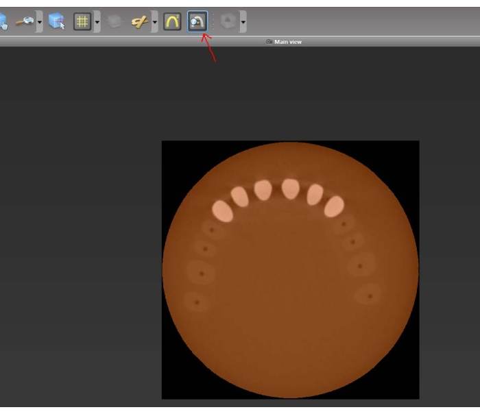
Figure 1: Measure the density of the teeth and the surrounding air. Average the measured values. (Arrow: density measuring tool). Please click here to view a larger version of this figure.
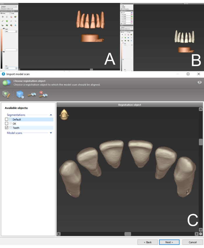
Figure 2: 3D reconstruction and segmentation. (A) 3D Reconstruction of pre-operative CBCT data. The lower threshold is adjusted to the calculated value. (B) Segmentation has been performed with the flood fill tool. The segmentation has been named "teeth" (color white). (C) Choose your segmentation as a registration object. Please click here to view a larger version of this figure.

Figure 3: Matching of CBCT and surface scan data. Check all the planes for correct alignment and finish the registration. Please click here to view a larger version of this figure.
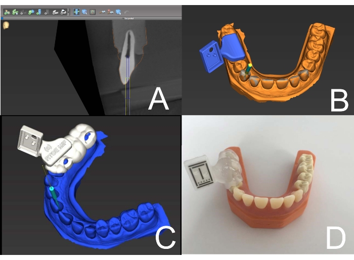
Figure 4: Access cavity planning and tray manufacturing. (A) The bur is virtually placed to the root canal orifice, providing straight-line access. (B) The marker tray is placed on the dental arch. (C) The marker tray has been designed to fit on the teeth surface. It is now ready to be exported and 3D printed. (D) The marker has been placed into the 3D-printed marker tray. Now the marker tray is placed on the dental arch and its fit is checked. Please click here to view a larger version of this figure.
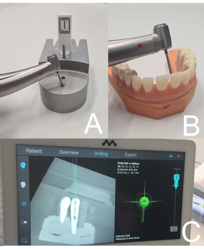
Figure 5: Bur registration and real-time visualization by the DNS. (A) Bur registration is performed with the associated tool. (B) Correct registration is checked before the treatment begins. The bur is placed to a prominent anatomic landmark (here incisal edge). The displayed position by the DNS should be exactly the same. (C) Display view of the DNS during access cavity preparation. Please click here to view a larger version of this figure.
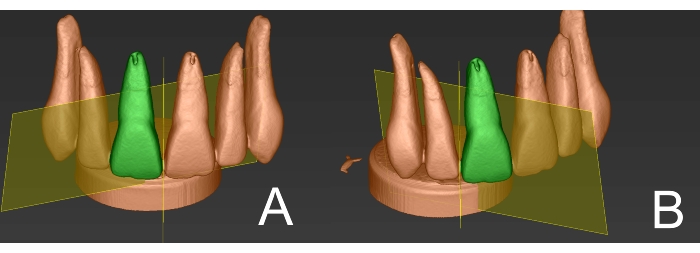
Figure 6: Single tooth segmentation for volume determination. (A) 3D reconstruction of CBCT data shows that teeth are connected due to proximal contacts. Two manual segmentation boundaries are drawn to provide a single tooth segmentation. Here: frontal view. (B) Lateral view. Please click here to view a larger version of this figure.

Figure 7: Matching of post- and pre-operative data. (A) Occlusal view of an endodontic access cavity that was performed with aid of a DNS. (B) Post-operative CBCT data in sagittal view. Note the straight-line access to the root canal space. (C) The post-operative segmentation of the tooth (red color) is matched with the pre-operative CBCT data (blue color). (D) 3D Models generated from the segmentation data are matched and show good accordance. Please click here to view a larger version of this figure.
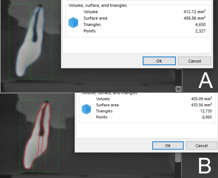
Figure 8: Volume calculation. (A) For the pre-operative 3D Model of the tooth, the planning software is able to calculate the volume in mm3. (B) Volume determination for the 3D Model of the tooth after access cavity preparation. Please click here to view a larger version of this figure.
Dyskusje
Several studies and case reports have demonstrated the feasibility of guided access cavity preparation in endodontics7. Navigation utilizing templates and sleeves for bur guidance (static navigation) was described to be a precise and safe method to access calcified root canals. Besides, the method was found to be independent from the operator's degree of clinical experience16, offering the possibility to treat teeth with advanced PCC without the risks of large loss of tooth structure or iatrogenic errors such as perforations.
When root canal treatment of posterior teeth with advanced PCC is indicated, static navigation utilizing templates and burs might become challenging due to the reduced interocclusal space, especially in patients with a reduced mouth opening7. A recent investigation revealed that deviations between planned and performed access cavities were significantly higher in molars compared to premolars or anterior teeth17, which was presumed to be attributed to interferences of the handpiece's head and the opposite teeth. A sleeveless template-based approach was described in a recent case report as an alternative to the mostly used sleeve-containing system and showed satisfying results18.
DNS provide real-time information about the spatial and angular deviation between the planned and the actual position of the bur that is used for access cavity preparation and thus there is no need for a template and its potentially reduced practicability in situations with reduced interocclusal space. Hence, DNS provide interoperative flexibility since the direction of access cavity preparation can be adjusted, which is not the case when a static navigation (template-based) approach is used.
Generally, the use of Guided Endodontics should be limited to teeth with advanced calcification, in which a conventional access cavity preparation is fraught with risk of iatrogenic errors, including root perforation and thus threatening tooth preservation, since the use of ionizing radiation (CBCT) is required for 3D planning. The use of CBCT in Endodontics should follow current scientific recommendations19. When generating the CBCT imaging data, a configuration with a limited field of view (FOV) will reduce the radiation dose. Visualization of highly calcified root canals can be enabled by a reduced voxel size, which allows accurate virtual 3D planning.
Also, the costs to perform a guided access cavity preparation are higher compared to the conventional technique. Until now, only a few DNS are available on the market, resulting in high acquisition fees. Nevertheless, static guided navigation also implies additional costs (template manufacturing process, sleeves, burs).
The results presented in the literature for the accuracy of DNS in non-surgical endodontic treatment are very promising. However, the few available systems consist of bulky and extraoral markers, which can reduce patient and operator comfort during the procedure. Here, the utilized DNS uses miniaturized components to avoid these disadvantages. Several studies in oral implantology20,21,22,23 and one investigation for endodontic access cavity preparation8 demonstrated the feasibility of this certain DNS and that it might become a potential alternative to template-based static navigation.
Sources for inaccuracies when using a DNS might potentially arise from planning errors. For example, full arch surface scans are still challenging24,25 for intraoral scanners and thus local deviations in the surface scan can occur and impair the precision of matching with the CBCT data.
For dynamic navigation also, the quality and fit of the marker tray is critical. Depending on the manufacturing process, material distortion26 might lead to deviations between the actual position and the displayed position of the bur. Geometrically considered, the deviation increases in case of a distortion when the angle between the camera and the marker is rather obtuse. Therefore, in the planning process for this specific DNS, it should be considered to place the marker tray in a position that provides a rather right angle between the camera and the marker surface. Nonetheless, in an in vitro study, there were no significant differences found between different types of marker positioning (contralateral/ipsilateral)23.
When performing volumetric measurements of pre- and post-operative conditions to determine the loss of tooth structure, it is crucial to use the same CBCT parameters and to set the same HU thresholds27. When a manual drawing of segmentation boundaries is necessary (in cases with proximal contacts) to perform a single tooth segmentation, inaccuracies might occur since the boundaries are drawn subjectively. More complex segmentation operations have been described in the literature to automatize the segmentation processes of teeth that have proximal contacts28,29. Nevertheless, inaccuracies due to manual segmentation boundaries in cases with proximal contacts are negligible in relation to the volume of substance loss.
Ujawnienia
All the authors declare that they have no conflicts of interest.
Podziękowania
None.
Materiały
| Name | Company | Catalog Number | Comments |
| Accuitomo 170 | Morita Manufacturing | NA | CBCT machine |
| coDiagnostiX | Dental Wings Inc | Version 10.4 | Planning software, which is mainly intended for implant surgery. Endodontic access cavities can be planned by adding the utlized bur to the implant database |
| DENACAM | mininavident | NA | Dynamic Nagivation System, consisting of (1) camera, which is mounted to an electric handpiece, (2) marker, (3)computer and screen, (4) associated software |
| TRIOS 3 | 3Shape A/S | NA | Surface scanner |
Odniesienia
- Patel, S., Rhodes, J. A practical guide to endodontic access cavity preparation in molar teeth. British Dental Journal. 203 (3), 133-140 (2007).
- Kiefner, P., Connert, T., ElAyouti, A., Weiger, R. Treatment of calcified root canals in elderly people: a clinical study about the accessibility, the time needed and the outcome with a three-year follow-up. Gerodontology. 34 (2), 164-170 (2017).
- Cvek, M., Granath, L., Lundberg, M. Failures and healing in endodontically treated non-vital anterior teeth with posttraumatically reduced pulpal lumen. Acta Odontologica Scandinavica. 40 (4), 223-228 (1982).
- Wigen, T. I., Agnalt, R., Jacobsen, I. Intrusive luxation of permanent incisors in Norwegians aged 6-17 years: a retrospective study of treatment and outcome. Dental Traumatology. 24 (6), 612-618 (2008).
- Andreasen, F. M., Zhijie, Y., Thomsen, B. L., Andersen, P. K. Occurrence of pulp canal obliteration after luxation injuries in the permanent dentition. Endodontics & Dental Traumatology. 3 (3), 103-115 (1987).
- Fleig, S., Attin, T., Jungbluth, H. Narrowing of the radicular pulp space in coronally restored teeth. Clinical Oral Investigations. 21 (4), 1251-1257 (2017).
- Moreno-Rabié, C., Torres, A., Lambrechts, P., Jacobs, R. Clinical applications, accuracy and limitations of guided endodontics: a systematic review. International Endodontic Journal. 53 (2), 214-231 (2020).
- Connert, T., et al. Real-time guided endodontics with a miniaturized dynamic navigation system versus conventional freehand endodontic access cavity preparation: substance loss and procedure time. Journal of Endodontics. 47 (10), 1651-1656 (2021).
- Zubizarreta-Macho, &. #. 1. 9. 3. ;., Muñoz, A. P., Deglow, E. R., Agustín-Panadero, R., Álvarez, J. M. Accuracy of computer-aided dynamic navigation compared to computer-aided static procedure for endodontic access cavities: An in vitro study. Journal of Clinical Medicine. 9 (1), 129 (2020).
- Jain, S. D., et al. Dynamically navigated versus freehand access cavity preparation: A comparative study on substance loss using simulated calcified canals. Journal of Endodontics. 46 (11), 1745-1751 (2020).
- Jain, S. D., Carrico, C. K., Bermanis, I. 3-Dimensional accuracy of dynamic navigation technology in locating calcified canals. Journal of Endodontics. 46 (6), 839-845 (2020).
- Gambarini, G., et al. Precision of dynamic navigation to perform endodontic ultraconservative access cavities: A preliminary in vitro analysis. Journal of Endodontics. 46 (9), 1286-1290 (2020).
- Dianat, O., et al. Accuracy and efficiency of a dynamic navigation system for locating calcified canals. Journal of Endodontics. 46 (11), 1719-1725 (2020).
- Dianat, O., Gupta, S., Price, J. B., Mostoufi, B. Guided endodontic access in a maxillary molar using a dynamic navigation system. Journal of Endodontics. 47 (4), 658-662 (2020).
- Chong, B. S., Dhesi, M., Makdissi, J. Computer-aided dynamic navigation: a novel method for guided endodontics. Quintessence International. 50 (3), 196-202 (2019).
- Connert, T., et al. Guided endodontics versus conventional access cavity preparation: A comparative study on substance loss using 3-dimensional-printed teeth. Journal of Endodontics. 45 (3), 327-331 (2019).
- Su, Y., et al. Guided endodontics: accuracy of access cavity preparation and discrimination of angular and linear deviation on canal accessing ability-an ex vivo study. BMC Oral Health. 21 (1), 606 (2021).
- Torres, A., Lerut, K., Lambrechts, P., Jacobs, R. Guided endodontics: Use of a sleeveless guide system on an upper premolar with pulp canal obliteration and apical periodontitis. Journal of Endodontics. 47 (1), 133-139 (2021).
- Patel, S., Brown, J., Semper, M., Abella, F., Mannocci, F. European Society of Endodontology position statement: Use of cone beam computed tomography in Endodontics: European Society of Endodontology (ESE) developed by. International Endodontic Journal. 52 (12), 1675-1678 (2019).
- Spille, J., et al. Comparison of implant placement accuracy in two different pre-operative digital workflows: navigated vs. pilot-drill-guided surgery. International Journal of Implant Dentistry. 7 (1), 1-9 (2021).
- Schnutenhaus, S., Knipper, A., Wetzel, M., Edelmann, C., Luthardt, R. Accuracy of computer-assisted dynamic navigation as a function of different intraoral reference systems: An In vitro study. International Journal of Environmental Research and Public Health. 18 (6), 3244 (2021).
- Edelmann, C., Wetzel, M., Knipper, A., Luthardt, R. G., Schnutenhaus, S. Accuracy of computer-assisted dynamic navigation in implant placement with a fully digital approach: A prospective clinical trial. Journal of Clinical Medicine. 10 (9), 1808 (2021).
- Duré, M., Berlinghoff, F., Kollmuss, M., Hickel, R., Huth, K. C. First comparison of a new dynamic navigation system and surgical guides for implantology: an in vitro study. International Journal of Computerized Dentistry. 24 (1), 9-17 (2021).
- Ender, A., Attin, T., Mehl, A. In vivo precision of conventional and digital methods of obtaining complete-arch dental impressions. Journal of Prosthetic Dentistry. 115 (3), 313-320 (2016).
- Ender, A., Zimmermann, M., Mehl, A. Accuracy of complete- and partial-arch impressions of actual intraoral scanning systems in vitro. International Journal of Computerized Dentistry. 22 (1), 11-19 (2019).
- Park, J. -. M., Jeon, J., Koak, J. -. Y., Kim, S. -. K., Heo, S. -. J. Dimensional accuracy and surface characteristics of 3D-printed dental casts. The Journal of Prosthetic Dentistry. 126 (3), 427-437 (2021).
- Dong, T., et al. Accuracy of in vitro mandibular volumetric measurements from CBCT of different voxel sizes with different segmentation threshold settings. BMC Oral Health. 19 (1), 206 (2019).
- Cui, Z., Li, C., Wang, W. ToothNet: automatic tooth instance segmentation and identification from cone beam CT images. Proceedings of the IEEE/CVF Conference on Computer Vision and Pattern Recognition (CVPR). , 6368-6377 (2019).
- Kim, S., Choi, S. Automatic tooth segmentation of dental mesh using a transverse plane). Annual International Conference of the IEEE Engineering in Medicine and Biology Society. Annual International Conference Journal. 2018, 4122-4125 (2018).
Przedruki i uprawnienia
Zapytaj o uprawnienia na użycie tekstu lub obrazów z tego artykułu JoVE
Zapytaj o uprawnieniaThis article has been published
Video Coming Soon
Copyright © 2025 MyJoVE Corporation. Wszelkie prawa zastrzeżone