Method Article
磷流式细胞仪与荧光细胞条形码用于单细胞信号分析和生物标志物发现
摘要
在这里, 提出了一种中-高通量分析在细胞级蛋白质磷酸化事件的协议。磷流式细胞术是表征信号畸变、识别和验证生物标志物、评估药效学的有力手段。
摘要
异常细胞信号在肿瘤的发展和进展中起着重要的作用。大多数新颖的靶向疗法确实是针对蛋白质和蛋白质功能, 细胞信号畸变可能因此作为标志物, 以表明个性化的治疗方案。与 DNA 和 RNA 分析相反, 蛋白质活动的变化可以更有效地评估药物敏感性和耐药性的机制。磷流式细胞术是一种功能强大的技术, 用于测量细胞水平的蛋白质磷酸化事件, 这是区别于其他基于抗体的方法的一个重要特征。该方法允许同时分析多种信号蛋白。结合荧光细胞条形码, 在短时间内, 标准细胞仪硬件可以获得较大的中、高吞吐量数据集。磷流式细胞术在基础生物学研究和临床研究中都有应用, 包括信号分析、生物标志物发现和药效学评价。以慢性淋巴细胞白血病细胞为例, 为纯化外周血单个核细胞磷流分析提供了详细的实验协议。
引言
磷流式细胞仪用于分析单细胞分辨率下蛋白质磷酸化水平。该方法的总体目标是在特定条件下映射细胞信号模式。利用流式细胞仪的多参数容量, 可以同时在不同的非均匀细胞群 (如外周血) 中同时分析几种信号通路。这些特性比其他基于抗体的技术具有优势, 如免疫组化、酶联免疫吸附试验 (ELISA)、蛋白质阵列和反相蛋白阵列 (RPPA)1。磷流式细胞术可与荧光细胞条形码 (FCB) 相结合, 这意味着单个细胞样本被标记为具有独特的荧光染料特征, 使它们可以混合在一起, 染色和分析为单个样品2。这降低了抗体的消耗, 提高了数据的鲁棒性, 通过组合控制和处理样本, 并提高了获取速度。组合 FCB 人口可以被分成较小的样本和染色多达35种不同的磷特定的抗体, 取决于数量的起始材料。因此, 大型分析实验可以使用标准的细胞仪硬件运行。磷流式细胞术应用于多例血液肿瘤患者样本中的特征信号通路, 包括慢性淋巴细胞白血病3、4、5、急性髓系白血病 (AML)6和非霍奇金淋巴瘤7。磷流式细胞术是一种有效的方法来表征信号畸变, 识别和验证生物标志物, 并评估药效学。
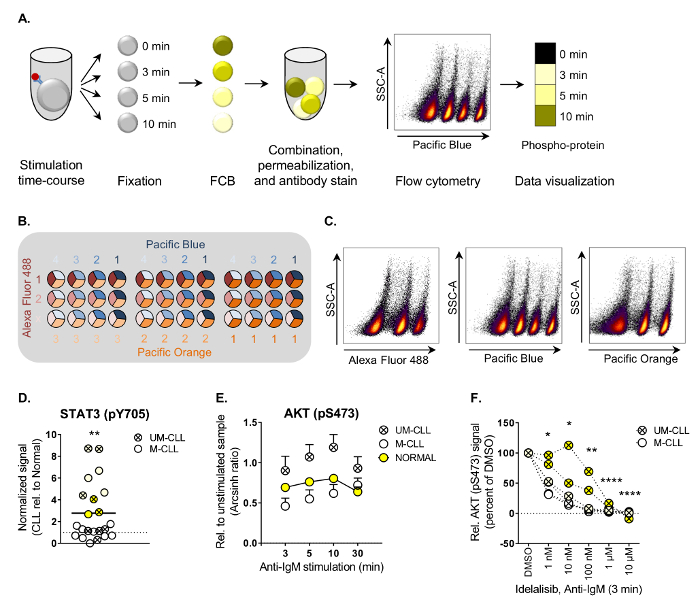
给出了磷流式细胞仪分析慢性淋巴细胞白血病患者标本的优化方案 (图 1A)。以基底信号特征、抗 IgM/B 细胞受体刺激和药物摄动为例。提供了一个 FCB 矩阵的详细描述。该协议可以很容易地适应其他悬浮细胞类型。
研究方案
根据所有捐助者的书面知情同意, 收到了血样。这项研究得到了挪威东南医学和健康研究伦理学区域委员会的批准, 并根据8赫尔辛基宣言进行了人体血液研究。
注:步骤1-3 应在无菌条件下进行组织培养罩。
1. 外周血单个核细胞 (PBMCs) 与慢血症患者血液样本的分离
注意: 人体血液应按照安全等级2的规定进行处理。
- 稀释血液1:1 与磷酸盐缓冲盐水 (PBS: 136.9 毫米氯化钠, 2.7 毫米氯化钾, 10.1毫米 Na2HPO4 x 2H2O, 1.8 毫米的 PO2, pH 4) 和转移到7.4 毫升管子 (50 毫升/管)。
- 小心10毫升的密度梯度介质 (例如, Lymphoprep) 到管底部使用10毫升吸管。
- 离心机在 800 x g 20 分钟在4摄氏度。PBMCs 现在可见于密度梯度介质层的顶端。
- 使用巴斯德吸管将细胞转化为两个新的50毫升管。用 PBS 洗两次 (填满管子)。
- 离心机在 350 x g 的15分钟, 丢弃上清和并用重悬3毫升的 PBS。
- 使用首选方法对单元格进行计数。
- 离心细胞在 350 x g 5 分钟丢弃上清。注:步骤1.8 到3.2 是可选的。可以直接进行步骤3.3。
- 并用重悬胎牛血清中的细胞, 辅以10% 二甲基亚砜 (亚砜), 并在适当的整除数中冷冻使用冷冻管。
注:亚砜对细胞是有毒的。一旦细胞与血清/亚砜混合, 就能快速工作。细胞可以长期储存在液氮中。
2. 细胞解冻
- 在37摄氏度的水浴中迅速解冻细胞。
注:亚砜对细胞是有毒的。快速工作以限制对亚砜的接触。 - 用10毫升的冷罗斯威尔公园纪念研究所培养基 (RPMI 1640 与补充 GlutaMAX, 见材料表) 洗涤细胞一次。
- 离心机以 300 x g 为5分钟, 丢弃上清液。
- 并用重悬 RPMI 1640 培养基中的细胞补充丙酮酸钠, 记忆非必需氨基酸和青霉素/链霉素 (根据指示加1x 稀释) 和10% 的血清。将细胞转移到小细胞培养瓶中, 在 5% CO2、37°c 1 小时内离开孵化器, 允许细胞校准。
3. 细胞的制备
- 使用首选方法计数可行的单元格。
- 将细胞转移到50毫升管和离心机在 300 x g 5 分钟。
- 并用重悬细胞在 RPMI 1640 培养基 (步骤 2.2) 补充1% 的血清不超过 50 x 106细胞/毫升。
- 将所需的细胞悬浮量转移到96井 V 型底板的井中。
注:井数对应于要测试的条件的数量。刺激时间的样本是从一个井中抽取的。计算50µL 样品每时间点 + 50 µL 的死容量。保存用于补偿控制的样本 (一个无瑕样本 + 每条形码染料一个样本)。 - 将96井板转移到预热的37°c 水浴中。把细胞休息10分钟。
4. 细胞的刺激和固定
注:在实验室的长凳上执行步骤 4-8 (即不育)。
注意: 固定缓冲 I 的主要成分是多聚甲醛, 这是有毒的 (吸入和皮肤接触)。小心处理。
- 准备一个96井 V 底板与60µL 的固定缓冲 I 每井样品。离开在37°c 水浴。
注:单元格: 修复缓冲区应为1:1。为了使蒸发在37摄氏度, 固定缓冲最初是丰富的。 - 或者, 在刺激前用药物治疗细胞。
- 将50µL 控制样品转移到固定板上。吹打上下混合。
- 或者, 启动刺激时间-路线通过增加10µg/毫升抗 IgM 细胞。吹打上下混合。
- 在每个时间点将50µL 样品转移到固定板上。吹打上下混合。
注:抗 IgM 诱导信号通常是早期启动的 (分钟)。 - 在添加最后一个样品后, 将固定板保持在37°c 10 分钟。
5. 荧光细胞条形码 (FCB)
注:见表 1列出的条形码试剂。
- 用 PBS 冲洗固定单元 3x (填好井)。
- 离心机以 500 x g 为5分钟, 丢弃上清液。
- 用条形码试剂制备96井 V 型底板。吸管5µL 每个条形码试剂每井的数量的组合所需的染色后的所有样品染色矩阵,如图 1B。每个样品将有一个独特的组合不同的条形码浓度。
- 并用重悬190µL 的细胞, 并转移到条形码板。完全混合。
注:染色一个补偿样品与最高的最终浓度用于每个条形码试剂和保存一个无瑕样品。 - 在室温下, 在黑暗中, 让细胞保持20分钟的温度。
- 用流动洗涤 (PBS, 1% 血清, 0.09% 叠氮化钠) 清洗染色的细胞 (填充水井)。
- 离心机以 500 x g 为5分钟, 丢弃上清液。
- 添加190µL 的流动洗涤细胞, 并结合条形码样品在一个15毫升管。将每个补偿控制转移到一个单独的1.7 毫升管。
- 离心机以 500 x g 为5分钟, 丢弃上清液。
6. 细胞通透胞内抗原染色
注意: 烫发缓冲 III 的主要成份是甲醇, 有毒 (吸入和皮肤接触) 和易燃。小心处理。
- 将2毫升的烫发缓冲器 III 转至15毫升管。留在摄氏-20 摄氏度, 所以它是冰后使用。
注:烫发缓冲器可以留在-20 摄氏度从实验开始。 - 添加1.5 毫升的冷烫发缓冲到条形码细胞 (在15毫升管) 和100µL 到每个补偿控制 (在1.7 毫升管) 下降明智的, 而涡流, 以避免细胞聚集在一起。
- 将细胞直接转移到-80 摄氏度。最少30分钟。
注:在这一点上暂停实验是很自然的。烫发缓冲器中的细胞可以长期储存在-80 摄氏度。
7. 抗体染色
注:请参阅资料表, 列出报告的磷抗体。
- 将细胞从-80 °c 转移到一盒冰上。
- 洗涤3x 与流动洗涤。
注:重要的是增加流洗涤, 以看到细胞颗粒,例如,增加3毫升的流动洗涤到条形码细胞的人口和1毫升的每个补偿控制。 - 离心机在 500 x g 5 分钟在4摄氏度。放弃上清。
- 并用重悬条形码细胞数量的流量洗涤, 这允许25µL 细胞悬浮每磷抗体染色。并用重悬200µL 流洗的补偿控制。
- 准备96井 V 底板染色抗体。最后的音量将是50µL/好。每一个井, 添加磷特定的抗体稀释在流动洗涤到最后的容量10µL, 表面标记稀释在流动洗涤到最后容量15µL 和25µL 细胞悬浮。
注:抗体稀释应在实验前滴定。包括同种控件。 - 在室温下, 在黑暗中, 让细胞保持30分钟的温度。
- 用流动洗涤 (填好井) 洗涤染色的细胞2x。
- 离心机以 500 x g 为5分钟, 丢弃上清液。
- 并用重悬细胞在150µL 的流动洗涤。
8. 编制补偿管制
- 在抗体染色的同时, 为抗体共轭荧光准备补偿控制。根据供应商的指示使用补偿珠。
9. 流式细胞仪分析
注:该实验可以运行在流式细胞仪与高通量取样器 (高温超导)。
- 利用无瑕控制优化光电倍增管 (PMT) 电压。
- 运行补偿控制并计算补偿矩阵。
- 运行示例。事件率应符合仪器规格。
10. 门控策略及数据分析
- 将 FCS 文件从实验中导入到流式细胞仪分析软件, 如 FlowJo 或 Cytobank (https://cellmass.cytobank.org)。
- 浇注策略
- 通过在密度点图中绘制 SSC-a与FSC-a 来选择淋巴细胞。
- 显示淋巴细胞和选择汗衫通过绘制 SSC与FSC-W。
- 通过绘制 SSC-A与曲面标记, 显示单个单元格并浇下单元格类型。
- 显示太平洋蓝与SSC 的细胞类型种群-密度图并根据其太平洋蓝染色强度选择不同的 FCB 种群 (见图 1A)。
- 在 FCB 通道上绘制磷抗体通道, 或作为热图 (参见图 1A) 显示磷酸化事件。
- 使用磷的逆双曲正弦 (arcsinh) (中值荧光强度) 计算磷信号与同种控制 (基底磷酸化水平, 见图 1D), 或刺激与静态细胞数量 (见图 1E)。
结果
磷流式细胞术协议的主要步骤如图 1A所示。在所提出的例子中, 条形码试剂蓝在四稀释染色的慢性淋巴细胞白血病细胞。三维条形码可以通过组合三条形码染料进行, 如图 1B所示。然后, 每个条形码试剂与SSC (图 1C) 上的后续浇口 deconvoluted 单个样本。表 1列出了有关条形码试剂的详细信息。
根据这里描述的过程, 磷蛋白水平的特点是 B 细胞的慢性淋巴细胞白血病患者和正常控制下的各种情况下3。对 B 细胞受体 (BCR) 下游20个信号分子的基底和刺激诱导磷酸化水平进行了分析 (见磷特定抗体列表的材料表)。根据正常对照组22例慢淋患者标本, 对基底磷蛋白水平进行了映射。分析表明, STAT3 (pY705) 在慢性淋巴细胞白血病细胞中明显上调 (图 1D)。STAT3 的本构活化在其他恶性血液病中有报道, 并与抗凋亡9有关。
为了识别通过 BCR 通路诱导的信号畸变, 细胞以抗 IgM 刺激, 可达30分钟。结果表明, IgVH unmutated 状态 (慢性淋巴细胞白血病) 患者的慢搏细胞显示对抗 IgM 刺激的敏感性提高10。这确实是观察到的大多数分析蛋白质, 但效果是统计学意义的, 只有 AKT (pS473) (图 1E, 嗯-慢白血病与 M 淋巴细胞白血病和正常)。为了检测异常 AKT (pS473) 信号是否可以逆转, 慢性淋巴细胞白血病细胞暴露于 PI3Kδ抑制剂 idelalisib, 用于治疗慢性淋巴细胞白血病患者11。如图 1F所示, AKT (pS473) 水平以浓度依赖性的方式在 idelalisib 治疗中明显减少, 表明激酶抑制剂可应用于慢性淋巴细胞化细胞异常信号的规范化。
这些结果表明, 磷流式细胞术与 FCB 结合是一种强有力的方法进行信号分析研究, 识别潜在的生物标志物, 并评估药效学。

图1。工作流程和应用磷流式细胞术分析的例子。
(A) 说明了磷流动程序的主要步骤。细胞首先被刺激, 然后被固定并且遭受 FCB, 在他们能被结合在一管为通透和随后抗体染色之后。细胞是运行在一个流动细胞仪, 并在数据分析过程中 deconvoluted 通过门。结果可以可视化为直方图或 heatmaps, 如图所示。(B) 使用 Alexa 氟 488 (三稀释)、太平洋蓝 (四稀释) 和太平洋橙 (三稀释) 的三维 FCB 染色基质的例子。这个矩阵将允许组合多达36个样本。(C) FCB 细胞的数量可以通过 deconvoluted 在每个 FCB 通道与SSC A 之间进行门控。分析软件中的门组合生成正确的分析种群。(D) 健康捐献者的静态 B 细胞 (n = 25) 和慢血症患者 (n = 22) 在 (A) 过程中经过磷流分析。以 IgGκ同种控制为 arcsinh 比, 计算了基底荧光强度信号。然后将 b 细胞中的信号规范化为 b 细胞中正常对照组的信号。**p < 0.01, 由不配对的两样本t检验计算。慢性淋巴细胞白血病: IgVH unmutated 慢性淋巴细胞白血病: IgVH 突变的慢性淋巴细胞白血病。相同颜色的符号代表患者样本, 根据20磷蛋白3的水平, 在分层集聚簇中组合在一起。(E) 正常控制下的 B 细胞 (n = 10, 平均 + sem) 或慢流病患者 (n = 11 [M-慢性淋巴细胞白血病] 和n = 8 [UM 淋巴细胞], 平均 + sem) 被刺激以抗 IgM 为指示的时间路线和受磷流动分析。对荧光强度信号进行了相对于静态样品的测量, 显示为 arcsinh 比。**p < 0.01 (正常 vs 慢性淋巴细胞) 和 **p < 0.001 (M-慢性淋巴细胞白血病), 用 Sidak 的多项比较试验计算。慢性淋巴细胞白血病: IgVH unmutated 慢性淋巴细胞白血病: IgVH 突变的慢性淋巴细胞白血病。(F) 在抗 IgM 刺激3分钟前, 用亚砜或 idelalisib 在20分钟内孵化出细胞。然后按照磷流协议处理这些单元格。*p < 0.05, **p < 0.01, ***p < 0.0001, 由 Sidak 的多次对比试验计算。慢性淋巴细胞白血病: IgVH unmutated 慢性淋巴细胞白血病: IgVH 突变的慢性淋巴细胞白血病。请参阅 (D) 解释符号颜色。(D F) 从3修改。请单击此处查看此图的较大版本.
| 系列稀释如下 (从库存解决方案开始) | ||||||
| 条形码试剂 | 库存浓度 | #1 | #2 | #3 | #4 | 无瑕 |
| Alexa 流体488 | 10毫克/毫升 | 1:500 | 1:5 | X | ||
| 太平洋蓝 | 10毫克/毫升 | 1:2500 | 1:4 | 1:4 | 1:10 | |
| 太平洋橙 | 2毫克/毫升 | 1:50 | 1:12 | 1:24 | ||
表 1.条形码试剂。
讨论
磷流式细胞术是测定单细胞蛋白质磷酸化水平的一种强有力的技术。由于该方法依赖于抗体的染色, 磷流式细胞术受抗体可用性的限制。此外, 为了获得可靠的结果, 所有抗体应在使用前滴定和验证。磷特定抗体的滴定详细的协议在别处被描述了12。在面板设计中, 考虑信噪比是至关重要的。在这个例子中, 所有的磷抗体被共轭到 Alexa 647。这种荧光通常提供低与高含量的磷蛋白样品之间的最佳差异。此外, 通过只使用一种颜色的磷蛋白质的其他渠道将是免费的 FCB 和表面标记染色。此面板设计减少了溢出到磷通道。通过将所有磷抗体共轭到同一荧光, 数据分析也将被简化。
在所提出的协议中, 所有的抗体染色在细胞的固定和通透后进行。然而, 重要的是要记住, 表面标记染色可能受到不利影响的固定和通透步骤, 由于表面抗原变性或增加非特异性染色13。因此, 使用者应在案件的基础上测试抗体的反应性。对兼容克隆的资源也可能有帮助, 例如对不同的固定/通透程序的概述以及它们与 https://www.cytobank.org/facselect/中各种抗体的相容性。
蛋白质磷酸化或脱磷酸是一种瞬变的改变, 发生在对外部和内在线索的反应。因此, 在比较磷酸化模式时, 必须在类似条件下进行实验。当研究主细胞从血液中发出信号时, 可能影响结果的因素包括在绘制血液、贮存条件以及在实验开始之前, 分离细胞的休息时间。当比较冷冻保存的细胞和新鲜分离的细胞从血液中的信号模式, 只有非常微小的显著差异可以观察到 (Skånland, 未发表)。但是, 例如, 在研究 biobanked 患者样本时, 仍宜使用冷冻保存正常细胞作为控制。磷流式细胞术实验的最佳条件和外部因素的影响应由个别用户进行测试。
本文给出了悬浮单元磷流分析的一种协议。该协议可以适应其他细胞类型, 但它是一个先决条件, 细胞作为单细胞悬浮在流式细胞术分析。实现这一目标的程序必须是微妙的, 以保持, 而不是影响, 磷酸化模式。例子存在黏附细胞被分离从培养的盘由冷的 trypsination12,14, 或者相当生长在微球15。当涉及到固体组织的磷流式细胞术, 一份报告存在于肺肿瘤, 其中单细胞通过通过一个管细胞过滤器16。最近, 磷流式细胞术结合了一种新的方法, 称为细胞内信号从组织 (解剖) 细胞分裂, 以研究磷蛋白在上皮组织17和大肠癌18。
FCB 是该议定书的一个关键步骤, 因为实验结束时样品的反褶积依赖于不同的 FCB 种群。为了得到这个, 细胞需要均匀染色。因此, 重要的是要准备一个条形码板, 细胞可以添加到。将试剂添加到细胞中会导致不均匀的染色和混合种群, 不能通过门 deconvoluted。强烈建议在实验执行之前对条形码稀释进行测试, 因为染色强度是与细胞类型相关的。
其他基于抗体的技术, 如蛋白质阵列和反相蛋白阵列 (RPPA) 可用于量化的磷蛋白水平, 在中等到高通量的方式。然而, 磷流式细胞术的一些性质将这种方法与其他的区别开来。磷流式细胞术的一个重要优点是它允许单细胞分析。通过将表面标记纳入不同的细胞子集, 可以检测出细胞间的异质性。此外, FCB 还允许对同一实验运行中的几个条件进行分析。这些特性使磷流式细胞术成为未来应用于生物标志物发现和精密医学19的一种诱人的方法。
披露声明
作者没有什么可透露的。
致谢
这项工作是在谢蒂尔·保尔森 Taskén 教授实验室进行的, 并得到了挪威癌症协会和 Stiftelsen 克里斯蒂安·卡尔森捷成的支持。约翰. Landskron 和玛丽安客运被承认对手稿的批判性阅读。
材料
| Name | Company | Catalog Number | Comments |
| RPMI 1640 GlutaMAX | ThermoFisher Scientific | 61870-010 | Cell culture medium |
| Fetal bovine serum | ThermoFisher Scientific | 10270169 | Additive to cell culture medium |
| Sodium pyruvate | ThermoFisher Scientific | 11360-039 | Additive to cell culture medium |
| MEM non-essential amino acids | ThermoFisher Scientific | 11140-035 | Additive to cell culture medium |
| Lymphoprep | Alere Technologies AS | 1114547 | Density gradient medium |
| Anti-IgM | Southern Biotech | 2022-01 | For stimulation of the B cell receptor |
| BD Phosflow Fix Buffer I | BD | 557870 | Fixation buffer |
| BD Phosflow Perm Buffer III | BD | 558050 | Permeabilization buffer |
| Alexa Fluor 488 5-TFP | ThermoFisher Scientific | A30005 | Barcoding reagent |
| Pacific Blue Succinimidyl Ester | ThermoFisher Scientific | P10163 | Barcoding reagent |
| Pacific Orange Succinimidyl Ester, Triethylammonium Salt | ThermoFisher Scientific | P30253 | Barcoding reagent |
| Compensation beads | Defined by user | Correct species reactivity | |
| Falcon tubes | Defined by user | ||
| Eppendorf tubes | Defined by user | ||
| 96 well V-bottom plates | Defined by user | Compatible with the flow cytometer | |
| Centrifuges | Defined by user | For Eppendorf tubes, Falcon tubes and plates | |
| Water bath | Defined by user | Temperature regulated | |
| Flow cytometer | Defined by user | With High Throughput Sampler (HTS) | |
| Name | Company | Catalog Number | Comments |
| Antigen | |||
| AKT (pS473) | Cell Signaling Technologies | 4075 | Clone: D9E Reference: Myhrvold et al., 2018, Single cell profiling of phospho-protein levels in.., Oncotarget, 9(10):9273-9284 Parente-Ribes et al., 2016, Spleen tyrosine kinase inhibitors reduce…, Haematologica, 101(2):e59-62 Skånland et al., 2014, T-cell co-stimulation through the CD2 and CD28…, Biochem J, 460(3):399-410 Kalland et al., 2012, Modulation of proximal signaling in normal and transformed…, Exp Cell Res, 318(14):1611-9 |
| ATF-2 (pT71) | Santa Cruz Biotechnology | sc-8398 | Clone: F-1 Reference: Skånland et al., 2014, T-cell co-stimulation through the CD2 and CD28…, Biochem J, 460(3):399-410 Pollheimer et al., 2013, Interleukin-33 drives a proinflammatory endothelial…, Arterioscler Thromb Vasc Biol, 33(2):e47-55 |
| BLNK (pY84) | Beckton Dickinson Pharmingen | 558443 | Clone: J117-1278 Reference: Myhrvold et al., 2018, Single cell profiling of phospho-protein levels in.., Oncotarget, 9(10):9273-9284 Parente-Ribes et al., 2016, Spleen tyrosine kinase inhibitors reduce…, Haematologica, 101(2):e59-62 Kalland et al., 2012, Modulation of proximal signaling in normal and transformed…, Exp Cell Res, 318(14):1611-9 Myklebust et al., 2017, Distinct patterns of B-cell receptor signaling in…, Blood, 129(6): 759-770 |
| Btk (pY223)/Itk (pY180) | Beckton Dickinson Pharmingen | 564846 | Clone: N35-86 Reference: Myklebust et al., 2017, Distinct patterns of B-cell receptor signaling in…, Blood, 129(6): 759-770 |
| Btk (pY551) | Beckton Dickinson Pharmingen | 558129 | Clone: 24a/BTK (Y551) Reference: Kalland et al., 2012, Modulation of proximal signaling in normal and transformed…, Exp Cell Res, 318(14):1611-9 |
| Btk (pY551)/Itk (pY511) | Beckton Dickinson Pharmingen | 558134 | Clone: 24a/BTK (Y551) Reference: Myhrvold et al., 2018, Single cell profiling of phospho-protein levels in.., Oncotarget, 9(10):9273-9284 Parente-Ribes et al., 2016, Spleen tyrosine kinase inhibitors reduce…, Haematologica, 101(2):e59-62 |
| CD3ζ (pY142) | Beckton Dickinson Pharmingen | 558489 | Clone: K25-407.69 Reference: Skånland et al., 2014, T-cell co-stimulation through the CD2 and CD28…, Biochem J, 460(3):399-410 |
| Histone H3 (pS10) | Cell Signaling Technologies | 9716 | Clone: D2C8 Reference: Myhrvold et al., 2018, Single cell profiling of phospho-protein levels in.., Oncotarget, 9(10):9273-9284 |
| IκBα | Cell Signaling Technologies | 5743 | Clone: L35A5 Reference: Myklebust et al., 2017, Distinct patterns of B-cell receptor signaling in…, Blood, 129(6): 759-770 |
| LAT (pY171) | Beckton Dickinson Pharmingen | 558518 | Clone: I58-1169 Reference: Skånland et al., 2014, T-cell co-stimulation through the CD2 and CD28…, Biochem J, 460(3):399-410 |
| Lck (pY505) | Beckton Dickinson Pharmingen | 558577 | Clone: 4/LCK-Y505 Reference: Myhrvold et al., 2018, Single cell profiling of phospho-protein levels in.., Oncotarget, 9(10):9273-9284 |
| MEK1 (pS298) | Beckton Dickinson Pharmingen | 560043 | Clone: J114-64 Reference: Myhrvold et al., 2018, Single cell profiling of phospho-protein levels in.., Oncotarget, 9(10):9273-9284 Skånland et al., 2014, T-cell co-stimulation through the CD2 and CD28…, Biochem J, 460(3):399-410 |
| NF-κB p65 (pS529) | Beckton Dickinson Pharmingen | 558422 | Clone: K10-895.12.50 Reference: Myhrvold et al., 2018, Single cell profiling of phospho-protein levels in.., Oncotarget, 9(10):9273-9284 Skånland et al., 2014, T-cell co-stimulation through the CD2 and CD28…, Biochem J, 460(3):399-410 Kalland et al., 2012, Modulation of proximal signaling in normal and transformed…, Exp Cell Res, 318(14):1611-9 Pollheimer et al., 2013, Interleukin-33 drives a proinflammatory endothelial…, Arterioscler Thromb Vasc Biol, 33(2):e47-55 |
| NF-κB p65 (pS536) | Cell Signaling Technologies | 4887 | Clone: 93H1 Reference: Myhrvold et al., 2018, Single cell profiling of phospho-protein levels in.., Oncotarget, 9(10):9273-9284 Skånland et al., 2014, T-cell co-stimulation through the CD2 and CD28…, Biochem J, 460(3):399-410 Kalland et al., 2012, Modulation of proximal signaling in normal and transformed…, Exp Cell Res, 318(14):1611-9 |
| p38 MAPK (pT180/Y182) | Cell Signaling Technologies | 4552 | Clone: 28B10 Reference: Myhrvold et al., 2018, Single cell profiling of phospho-protein levels in.., Oncotarget, 9(10):9273-9284 Skånland et al., 2014, T-cell co-stimulation through the CD2 and CD28…, Biochem J, 460(3):399-410 Pollheimer et al., 2013, Interleukin-33 drives a proinflammatory endothelial…, Arterioscler Thromb Vasc Biol, 33(2):e47-55 |
| p44/42 MAPK (pT202/Y204) | Cell Signaling Technologies | 4375 | Clone: E10 Reference: Myhrvold et al., 2018, Single cell profiling of phospho-protein levels in.., Oncotarget, 9(10):9273-9284 Parente-Ribes et al., 2016, Spleen tyrosine kinase inhibitors reduce…, Haematologica, 101(2):e59-62 Skånland et al., 2014, T-cell co-stimulation through the CD2 and CD28…, Biochem J, 460(3):399-410 Kalland et al., 2012, Modulation of proximal signaling in normal and transformed…, Exp Cell Res, 318(14):1611-9 Pollheimer et al., 2013, Interleukin-33 drives a proinflammatory endothelial…, Arterioscler Thromb Vasc Biol, 33(2):e47-55 |
| p53 (pS15) | Cell Signaling Technologies | NN | Clone: 16G8 Reference: Irish et al., 2007, Flt3 Y591 duplication and Bcl-2 overexpression…, Blood, 109(6):2589-96 |
| p53 (pS20) | Cell Signaling Technologies | NN | Clone: Polyclonal Reference: Irish et al., 2007, Flt3 Y591 duplication and Bcl-2 overexpression…, Blood, 109(6):2589-96 |
| p53 (pS37) | Cell Signaling Technologies | NN | Clone: Polyclonal Reference: Irish et al., 2007, Flt3 Y591 duplication and Bcl-2 overexpression…, Blood, 109(6):2589-96 |
| p53 (pS46) | Cell Signaling Technologies | NN | Clone: Polyclonal Reference: Irish et al., 2007, Flt3 Y591 duplication and Bcl-2 overexpression…, Blood, 109(6):2589-96 |
| p53 (pS392) | Cell Signaling Technologies | NN | Clone: Polyclonal Reference: Irish et al., 2007, Flt3 Y591 duplication and Bcl-2 overexpression…, Blood, 109(6):2589-96 |
| PLCγ2 (pY759) | Beckton Dickinson Pharmingen | 558498 | Clone: K86-689.37 Reference: Myhrvold et al., 2018, Single cell profiling of phospho-protein levels in.., Oncotarget, 9(10):9273-9284 Myklebust et al., 2017, Distinct patterns of B-cell receptor signaling in…, Blood, 129(6): 759-770 |
| Rb (pS807/pS811) | Beckton Dickinson Pharmingen | 558590 | Clone: J112-906 Reference: Myhrvold et al., 2018, Single cell profiling of phospho-protein levels in.., Oncotarget, 9(10):9273-9284 Pollheimer et al., 2013, Interleukin-33 drives a proinflammatory endothelial…, Arterioscler Thromb Vasc Biol, 33(2):e47-55 |
| S6-Ribos. Prot. (pS235/236) | Cell Signaling Technologies | 4851 | Clone: D57.2.2E Reference: Myhrvold et al., 2018, Single cell profiling of phospho-protein levels in.., Oncotarget, 9(10):9273-9284 |
| SAPK/JNK (pT183/Y185) | Cell Signaling Technologies | 9257 | Clone: G9 Reference: Myhrvold et al., 2018, Single cell profiling of phospho-protein levels in.., Oncotarget, 9(10):9273-9284 Pollheimer et al., 2013, Interleukin-33 drives a proinflammatory endothelial…, Arterioscler Thromb Vasc Biol, 33(2):e47-55 |
| SLP76 (pY128) | Beckton Dickinson Pharmingen | 558438 | Clone: J141-668.36.58 Reference: Skånland et al., 2014, T-cell co-stimulation through the CD2 and CD28…, Biochem J, 460(3):399-410 |
| STAT1 (pY701) | Beckton Dickinson Pharmingen | 612597 | Clone: 4a Reference: Myhrvold et al., 2018, Single cell profiling of phospho-protein levels in.., Oncotarget, 9(10):9273-9284 Myklebust et al., 2017, Distinct patterns of B-cell receptor signaling in…, Blood, 129(6): 759-770 |
| STAT3 (pY705) | Beckton Dickinson Pharmingen | 557815 | Clone: 4/P-STAT3 Reference: Myhrvold et al., 2018, Single cell profiling of phospho-protein levels in.., Oncotarget, 9(10):9273-9284 |
| STAT4 (pY693) | Zymed/ThermoFisher Scientific | 71-7900 | Clone: Polyclonal Reference: Uzel et al., 2001, Detection of intracellular phosphorylated STAT-4 by flow cytometry, Clin Immunol, 100(3): 270-6 |
| STAT5 (pY694) | Beckton Dickinson Pharmingen | 612599 | Clone: 47/Stat5(pY694) Reference: Myhrvold et al., 2018, Single cell profiling of phospho-protein levels in.., Oncotarget, 9(10):9273-9284 Skånland et al., 2014, T-cell co-stimulation through the CD2 and CD28…, Biochem J, 460(3):399-410 Myklebust et al., 2017, Distinct patterns of B-cell receptor signaling in…, Blood, 129(6): 759-770 |
| STAT6 (pY641) | Beckton Dickinson Pharmingen | 612601 | Clone: 18/P-Stat6 Reference: Myhrvold et al., 2018, Single cell profiling of phospho-protein levels in.., Oncotarget, 9(10):9273-9284 |
| SYK (pY525/Y526) | Cell Signaling Technologies | 12081 | Clone: C87C1 Reference: Myhrvold et al., 2018, Single cell profiling of phospho-protein levels in.., Oncotarget, 9(10):9273-9284 Parente-Ribes et al., 2016, Spleen tyrosine kinase inhibitors reduce…, Haematologica, 101(2):e59-62 |
| ZAP70/SYK (pY319/Y352) | Beckton Dickinson Pharmingen | 557817 | Clone: 17A/P-ZAP70 Reference: Skånland et al., 2014, T-cell co-stimulation through the CD2 and CD28…, Biochem J, 460(3):399-410 Kalland et al., 2012, Modulation of proximal signaling in normal and transformed…, Exp Cell Res, 318(14):1611-9 Myklebust et al., 2017, Distinct patterns of B-cell receptor signaling in…, Blood, 129(6): 759-770 |
参考文献
- Lu, Y., et al. Using reverse-phase protein arrays as pharmacodynamic assays for functional proteomics, biomarker discovery, and drug development in cancer. Seminars in Oncology. 43 (4), 476-483 (2016).
- Krutzik, P. O., Nolan, G. P. Fluorescent cell barcoding in flow cytometry allows high-throughput drug screening and signaling profiling. Nature Methods. 3 (5), 361-368 (2006).
- Myhrvold, I. K., et al. Single cell profiling of phospho-protein levels in chronic lymphocytic leukemia. Oncotarget. 9 (10), 9273-9284 (2018).
- Parente-Ribes, A., et al. Spleen tyrosine kinase inhibitors reduce CD40L-induced proliferation of chronic lymphocytic leukemia cells but not normal B cells. Haematologica. 101 (2), e59-e62 (2016).
- Blix, E. S., et al. Phospho-specific flow cytometry identifies aberrant signaling in indolent B-cell lymphoma. BMC Cancer. 12, 478(2012).
- Irish, J. M., et al. Single cell profiling of potentiated phospho-protein networks in cancer cells. Cell. 118 (2), 217-228 (2004).
- Myklebust, J. H., et al. Distinct patterns of B-cell receptor signaling in non-Hodgkin lymphomas identified by single-cell profiling. Blood. 129 (6), 759-770 (2017).
- World Medical Association. World Medical Association Declaration of Helsinki: ethical principles for medical research involving human subjects. THE JOURNAL OF THE AMERICAN MEDICAL ASSOCIATION. 310 (20), 2191-2194 (2013).
- Siveen, K. S., et al. Targeting the STAT3 signaling pathway in cancer: role of synthetic and natural inhibitors. Biochimica et Biophysica Acta. 1845 (2), 136-154 (2014).
- Fabbri, G., Dalla-Favera, R. The molecular pathogenesis of chronic lymphocytic leukaemia. Nature Reviews Cancer. 16 (3), 145-162 (2016).
- Arnason, J. E., Brown, J. R. Targeting B Cell Signaling in Chronic Lymphocytic Leukemia. Current Oncology Reports. 19 (9), 61(2017).
- Landskron, J., Tasken, K. Phosphoprotein Detection by High-Throughput Flow Cytometry. Methods in Molecular Biology. 1355, 275-290 (2016).
- Krutzik, P. O., Clutter, M. R., Nolan, G. P. Coordinate analysis of murine immune cell surface markers and intracellular phosphoproteins by flow cytometry. Journal of Immunology. 175 (4), 2357-2365 (2005).
- Pollheimer, J., et al. Interleukin-33 drives a proinflammatory endothelial activation that selectively targets nonquiescent cells. Arteriosclerosis, Thrombosis, and Vascular Biology. 33 (2), e47-e55 (2013).
- Ertsås, H. C., Nolan, G. P., LaBarge, M. A., Lorens, J. B. Microsphere cytometry to interrogate microenvironment-dependent cell signaling. Integrative biology: quantitative biosciences from nano to macro. 9 (2), 123-134 (2017).
- Lin, C. C., et al. Single cell phospho-specific flow cytometry can detect dynamic changes of phospho-Stat1 level in lung cancer cells. Cytometry A. 77 (11), 1008-1019 (2010).
- Simmons, A. J., et al. Cytometry-based single-cell analysis of intact epithelial signaling reveals MAPK activation divergent from TNF-alpha-induced apoptosis in vivo. Molecular Systems Biology. 11 (10), 835(2015).
- Simmons, A. J., et al. Impaired coordination between signaling pathways is revealed in human colorectal cancer using single-cell mass cytometry of archival tissue blocks. Science Signaling. 9 (449), rs11(2016).
- Friedman, A. A., Letai, A., Fisher, D. E., Flaherty, K. T. Precision medicine for cancer with next-generation functional diagnostics. Nature Reviews Cancer. 15 (12), 747-756 (2015).
转载和许可
请求许可使用此 JoVE 文章的文本或图形
请求许可This article has been published
Video Coming Soon
版权所属 © 2025 MyJoVE 公司版权所有,本公司不涉及任何医疗业务和医疗服务。
我们使用 cookie 来增强您在我们网站上的体验。
继续使用我们的网站或单击“继续”,即表示您同意接受我们的 cookie。