Using Diffusion Tensor Imaging in Traumatic Brain Injury
Genel Bakış
Source: Laboratories of Jonas T. Kaplan and Sarah I. Gimbel—University of Southern California
Traditional brain imaging techniques using MRI are very good at visualizing the gross structures of the brain. A structural brain image made with MRI provides high contrast of the borders between gray and white matter, and information about the size and shape of brain structures. However, these images do not detail the underlying structure and integrity of white matter networks in the brain, which consist of axon bundles that interconnect local and distant brain regions.
Diffusion MRI uses pulse sequences that are sensitive to the diffusion of water molecules. By measuring the direction of diffusion, it is possible to make inferences about the structure of white matter networks in the brain. Water molecules within an axon are constrained in their movements by the cell membrane; instead of randomly moving in every direction with equal probability (isotropic movement), they are more likely to move in certain directions, in parallel with the axon (anisotropic movement; Figure 1). Therefore, measures of diffusion anisotropy are thought to reflect properties of the white matter such as fiber density, axon thickness, and degree of myelination. One common measure is fractional anisotropy (FA). FA values range from 0, which represents completely isotropic movement, to 1, reflecting maximum anisotropy.
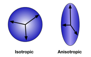
Figure 1: Diffusion anisotropy. When the direction of diffusion is unconstrained and random, movement is measured in all directions equally. This is isotropic diffusion (A). When water molecules are contained within the axon of a neuron, diffusion is anisotropic, tending to occur more frequently along the direction of the axon (B). Please click here to view a larger version of this figure.
In this experiment we will use diffusion tensor imaging (DTI) to measure white matter integrity in traumatic brain injury (TBI). TBI occurs when an external force injures the brain, such as a blow to the head or a sudden movement like the kind that might occur in a car accident. This type of brain injury from mechanical forces is associated with diffuse axonal injury-damage to white matter throughout the brain. Because it is an injury affecting white matter integrity, standard neuroimaging techniques may not reveal the damage. However, measures of diffusion are especially sensitive to these anatomical changes. Following a study by Kraus et al.1, we compare a group of healthy controls to a group of people with TBI and use diffusion imaging to measure the effect of TBI on cerebral white matter. Furthermore, we will test the relationship between white matter integrity and cognitive function using an attention task.2 This study uses a region of interest (ROI) approach focusing on three white matter tracts: the splenium of the corpus callosum, the anterior corona radiata, and the superior longitudinal fasciculus (Figure 2).
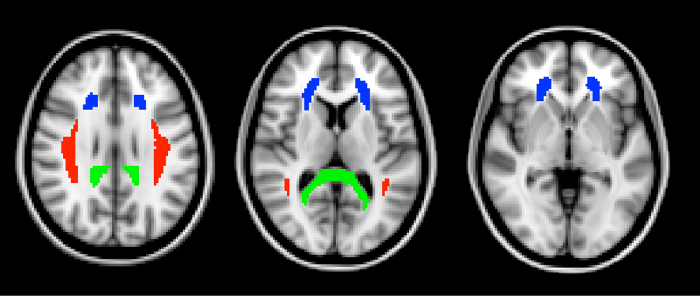
Figure 2: Regions of interest. The three ROIs, defined from the ICBM DTI-81 atlas, are shown here in horizontal slices through the brain. In green is the splenium of the corpus callosum. The splenium is the most posterior part of the corpus callosum. In blue is the anterior corona radiata. The superior longitudinal fasciculus is shown in red. Please click here to view a larger version of this figure.
Prosedür
1. Participant recruitment
- Recruit 20 participants with moderate to severe TBI, and 20 age-matched controls. All participants should be over the age of 18.
- TBI patients should have experienced a closed head injury that occurred at least 6 months ago. TBI is diagnosed by assessing several factors such as changes in consciousness, loss of consciousness, and loss of memory from before or after the accident. To be classified as moderate to severe, the patient must have experienced a period of loss of consciousness that was greater than 30 min, and/or obtained a score of less than 13 on the Glasgow Coma Scale.
- Control participants should have no history of neurological or psychological disorders.
- All participants should not have metal in their body. This is an important safety requirement due to the high magnetic field involved in MRI.
- All participants should not suffer from claustrophobia, since the MRI requires lying in the small space of the scanner bore.
- Pre-scan procedures
- Fill out pre-scan paperwork.
- When participants come in for their MRI scan, have them first fill out a metal screen form to make sure they have no counter-indications for MRI, an incidental findings form giving consent for their scan to be looked at by a radiologist, and a consent form detailing the risks and benefits of the study.
- Prepare participants to go in the scanner by removing all metal from their body, including belts, wallets, phones, hair clips, coins, and all jewelry.
- Scanner preparation
- Give the participant ear plugs to protect their ears from the noise of the scanner and ear phones to wear so they can hear the experimenter during the scan, and have them lie down on the bed with their head in the coil.
- Give the participant the emergency squeeze ball and instruct them to squeeze it in case of emergency during the scan.
- Use foam pads to secure the participants head in the coil to avoid excess movement during the scan, and remind the participant that it is very important to stay as still as possible during the scan, as even the smallest movements blur the images.
- Data collection
- Collect a high-resolution T1-weighted anatomical scan. This will be used for registering the participant's brain to the standard atlas space.
- Begin scanning using a pulse sequence optimized for DTI.
- One B0 image is acquired that is not sensitive to diffusion direction.
- Multiple diffusion-weighted images are acquired, each sensitive to a different direction of diffusion. The more directions acquired, the more finely we will be able to resolve the diffusion tensor. However, increasing the number of directions also increases acquisition time. In this study, we will acquire 64 different directions.
- Attention task
- Outside the MRI scanner, have all participants perform a version of the Attention Network Task (ANT)3 to assess their selective attention ability.
- Seat the participant in front of a computer screen, and instruct them on how to complete the task.
- Explain that a series of arrows will appear on the screen. The participant's task is to respond only to the arrow in the center, and to ignore the others. If the central arrow is pointing left, they will press the 'F' key with their left hand. If the central arrow is pointing right, they will press the 'J' key with their right hand. They should respond as quickly and as accurately as possible.
- Begin the task.
- On each trial, present a row of five arrows on the screen. Each arrow can be pointing left or right. On congruent trials, all of the arrows point in the same direction. On incongruent trials, the center arrow points in the opposite direction from the flanking arrows. Each trial begins with a fixation cross that remains on the screen for a variable duration between 400 and 1600 ms. Then the arrow stimuli appear and remain on the screen until the participant responds, or for a maximum of 1700 ms. The trial concludes with a fixation cross that remains on the screen until a total trial duration of 4s has been reached.
- Present 100 trials, half with congruent targets and half incongruent targets.
- Compute the difference in reaction time between incongruent targets and congruent targets. Typically, reaction time is slower in response to incongruent targets. People who are more distracted by the flanking arrows will have a larger difference in reaction time between incongruent and congruent targets. This measure of attentional control will be tested against measures of white matter integrity.
- Post-experiment procedures
- Debrief the participant.
- Pay the participant.
- Data analysis
- Pre-process the diffusion data.
- Visually inspect the data to make sure that it is free from artifacts.
- Perform eddy-current correction with specialized software.
- For each subject, register each of the directional diffusion images to the B0 image using a linear rigid-body affine transformation. This step will compensate for any motion that occurred from scan to scan.
- Remove the skull and other non-brain tissues from the images using automated software. This will ensure that we do not calculate tensors for voxels that are outside the brain.
- Combine among the multiple direction images to calculate the diffusion tensor at each voxel. There are several freely available software packages for processing DTI data that will calculate these values.
- Calculate FA at each voxel, the proportion of tensor magnitude due to anisotropic diffusion.
- Register the diffusion images to the high-resolution anatomical T1 image, and then to the standard atlas space to allow for group-level analysis.
- Define regions of interest (ROI).
- Obtain the three ROI masks from a standard white matter atlas. Here, we use the ICBM-DTI-81 white matter atlas created by the International Consortium for Brain Mapping (Figure 2).
- Register each individual subject's high-resolution anatomical image to the standard atlas.
- Warp the ROI masks into each participant's individual brain space using the registrations performed in the previous step.
- Extract FA values for each subject from each of the three ROIs.
- Compare the FA values between the two groups using Analysis of Variance (ANOVA).
- Compute the Pearson correlation between participants' congruency scores from the ANT and the FA values.
- Pre-process the diffusion data.
Sonuçlar
The FA values from the three ROIs are shown in Figure 3. Fractional anisotropy was significantly lower in the TBI group in all three ROIs, indicating the presence of widespread white matter damage in those individuals. This non-localized loss of white matter integrity is typical of TBI.
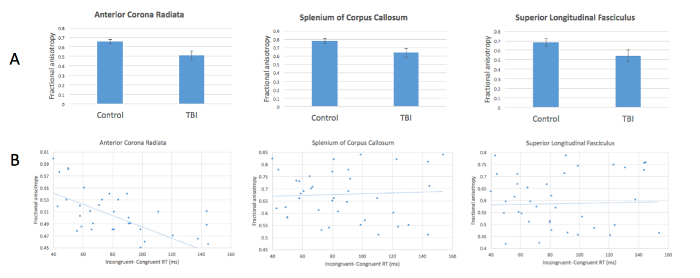
Figure 3: Reduced anisotropy in patients with TBI and relationship with attentional control. (A) FA values are significantly lower in TBI patients compared with healthy controls in all 3 ROIs. (B) FA in the anterior corona radiata correlates negatively with increased incongruency effect in the attention task. Please click here to view a larger version of this figure.
Our measure of attentional control-response time differences between congruent and incongruent targets-correlated negatively with FA values in the anterior corona radiata (Figure 3). In other words, greater differences in response time, indicating poorer attentional control, are associated with decreased FA. These results evidence a relationship between white matter integrity in this location and performance on this task. This relationship was not found in the other two ROIs. The anterior corona radiata is associated with connections to the anterior cingulate cortex, a structure known to play an important part in attentional control.
Başvuru ve Özet
Because diffusion imaging can reveal differences in white matter structure that are often not visible with traditional MRI imaging, it is an important tool for understanding brain structure and function. In this experiment we identified a clinically relevant marker for traumatic brain injury that may be used to predict the behavioral consequences of such an injury. DTI has been especially useful in the study of brain development, as changes in white matter structure are found throughout the lifespan from early childhood through late adulthood. For example, aging in older adults is associated with a decline in fractional anisotropy.
More sophisticated analysis of diffusion images allows for the reconstruction and tracing of fiber tracts in the brain, a process known as tractography. Tractography uses the directional information in contiguous voxels to trace specific fiber bundles as they traverse through the brain and can help to build models of the various interconnections among brain structures. This technique can be used to study the connections between individual brain regions of interest, or alternatively to analyze the entire connectome, or complex network structure, of the brain.
Referanslar
- Kraus, M.F., et al. White matter integrity and cognition in chronic traumatic brain injury: a diffusion tensor imaging study. Brain. 130, 2508-2519 (2007).
- Niogi, S.N., et al. Structural dissociation of attentional control and memory in adults with and without mild traumatic brain injury. Brain. 131, 3209-3221 (2008).
- Fan, J., McCandliss, B.D., Sommer, T., Raz, A., & Posner, M.I. Testing the efficiency and independence of attentional networks. J Cogn Neurosci. 14, 340-347 (2002).
Etiketler
Atla...
Bu koleksiyondaki videolar:

Now Playing
Using Diffusion Tensor Imaging in Traumatic Brain Injury
Neuropsychology
16.7K Görüntüleme Sayısı

The Split Brain
Neuropsychology
68.2K Görüntüleme Sayısı

Motor Maps
Neuropsychology
27.4K Görüntüleme Sayısı

Perspectives on Neuropsychology
Neuropsychology
12.0K Görüntüleme Sayısı

Decision-making and the Iowa Gambling Task
Neuropsychology
32.3K Görüntüleme Sayısı

Executive Function in Autism Spectrum Disorder
Neuropsychology
17.6K Görüntüleme Sayısı

Anterograde Amnesia
Neuropsychology
30.3K Görüntüleme Sayısı

Physiological Correlates of Emotion Recognition
Neuropsychology
16.2K Görüntüleme Sayısı

Event-related Potentials and the Oddball Task
Neuropsychology
27.4K Görüntüleme Sayısı

Language: The N400 in Semantic Incongruity
Neuropsychology
19.5K Görüntüleme Sayısı

Learning and Memory: The Remember-Know Task
Neuropsychology
17.1K Görüntüleme Sayısı

Measuring Grey Matter Differences with Voxel-based Morphometry: The Musical Brain
Neuropsychology
17.3K Görüntüleme Sayısı

Decoding Auditory Imagery with Multivoxel Pattern Analysis
Neuropsychology
6.4K Görüntüleme Sayısı

Visual Attention: fMRI Investigation of Object-based Attentional Control
Neuropsychology
41.5K Görüntüleme Sayısı

Using TMS to Measure Motor Excitability During Action Observation
Neuropsychology
10.1K Görüntüleme Sayısı
JoVE Hakkında
Telif Hakkı © 2020 MyJove Corporation. Tüm hakları saklıdır