
Photoacoustic Tomography to Image Blood and Lipids in the Infrarenal Aorta
Overzicht
Source: Gurneet S. Sangha and Craig J. Goergen, Weldon School of Biomedical Engineering, Purdue University, West Lafayette, Indiana
Photoacoustic tomography (PAT) is an emerging biomedical imaging modality that utilizes light generated acoustic waves to obtain compositional information from tissue. PAT can be used to image blood and lipid components, which is useful for a wide variety of applications, including cardiovascular and tumor imaging. Currently used imaging techniques have inherent limitations that restrict their use with researchers and physicians. For example, long acquisition times, high costs, use of harmful contrast, and minimal to high invasiveness are all factors that limit the use of various modalities in the laboratory and clinic. Currently, the only comparable imaging techniques to PAT are emerging optical techniques. But these also have disadvantages, such as limited depth of penetration and the need for exogenous contrast agents. PAT provides meaningful information in a rapid, noninvasive, label-free manner. When coupled with ultrasound, PAT can be used to obtain structural, hemodynamic, and compositional information from tissue, thereby complementing currently used imaging techniques. The advantages of PAT illustrate its capabilities to make an impact in both the preclinical and clinical environment.
Principes
PAT is a hybrid modality that utilizes light-induced acoustic waves to obtain compositional information from tissue. Acoustic propagation is attributed to thermoelastic expansion. This occurs when specific chemical bonds in a tissue absorb light, and the ambient temperature rise causes the tissue to expand. To elaborate, specific chemical bonds absorb light, causing the molecule to vibrate and convert some of this vibrational energy to heat. This production of heat causes local tissue expansion, which induces acoustic propagations that can be detected by an ultrasound transducer. To induce the photoacoustic effect, both the thermal and stress confinement conditions must be met to minimize heat dissipation and allow thermoelastically-induced pressure to build up within tissue. The resulting photoacoustic pressure wave can be characterized by equation (1), which states that the light-induced acoustic wave (Po) is governed by the temperature-dependent Grueneisen parameter (Γ), absorption coefficient (µa), and local optical fluence (F).
Po = ΓµaF Equation 1
As a result, each mK rise in temperature characteristically produces a 800 Pascal pressure wave that can be detected using an ultrasound transducer. This bond-selective absorption of light allows users to target various biological components by tuning the wavelength of light such as using 1100 nm light to target blood and 1210 nm light to target lipids. Additionally, since light is being used to induce acoustic wave propagation, this technique can be used to typically image deeper structures than other optical techniques without the need for contrast agents or invasive procedures. This specific method using long-wavelength light in the second near-infrared window light to induce acoustic waves provides numerous advantages to the user, allowing vibrational PAT (or VPAT) to be potentially used for a wide range of biomedical applications.
Procedure
The following procedure describes the methods needed to set up VPAT for blood and lipid imaging of the infrarenal aorta in apolipoprotein-E deficient (apoE-/-) mice.
1. Laser-ultrasound Coupling
- Obtain a Nd:YAG pulsed optical parametric oscillator laser and an ultrasound system. Acquire a pulse generator, 1 BNC cable, and a D connector attached to two BNC cables.
- Using the D connector set-up, attach the 'Fire' BNC cable to port A of the pulse generator, and 'Q-switch' to port B of the pulse generator. Finally, attach a BNC cable from port C to 'trigger in' on the back of the ultrasound system.
- Align the fiber optic cable with the laser and attach the fiber ends to the sides of the 40 MHz ultrasound transducer.
- Adjust the delay of ports A, B, and C to the values listed here (Port A: 0.00000000, Port B: 0.00021440, Port C: 0.00000910). Set ports A and B to inverted signal and port C to normal signal.
2. Animal Preparation and Image Acquisition
- Anesthetize an apoE-/- mouse using 3% isoflurane in an anesthesia induction chamber. Once the animal is anesthetized move the mouse to a nose cone to deliver 1-2% isoflurane.
- Apply eye lubricate to the animals eyes to prevent corneal desiccation. Tape the mouse's paws to electrodes built into the heated stage to monitor animal's respiration and heart rate. Finally, insert rectal probe to monitor body temperature.
- Apply depilatory cream to remove hair from the animal's abdomen. Wipe off after 30 s with a gauze pad.
- Place the ultrasound transducer on the animal's abdomen and locate the infrarenal aorta. The left renal vein and the aortic trifurcation into the tail artery are two landmarks that will help the user locate this area.
- Run the laser to output 1100 nm light to target blood followed by 1210 nm light to target lipid. Use appropriate laser safety goggles when the laser is in use.
Resultaten
Here, VPAT methods were used to perform lipid and blood specific imaging in vivo. By coupling a laser and ultrasound system, light was delivered to tissue and the resulting acoustic waves were detected. Ultrasound imaging allowed us to obtain structural information of the infrarenal aorta (Figure 1a) that can be used to better interpret VPAT compositional information. Specifically, a 1100 nm light was used to image blood within the aorta (Figure 1b), and a 1210 nm light was used to image subcutaneous and periaortic fat accumulation (Figure 1c). From the ultrasound and VPAT images, one can see that the subcutaneous fat follows the geometry of the skin, the periaortic fat follows the contour of the aorta, and the blood signal originates from within the aorta. These results confirm that, indeed, VPAT can be used to image blood and lipid accumulation in vivo.
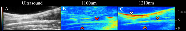
Figure 1: Ultrasound (left), blood VPAT (middle), and lipid VPAT (right) images of ApoE-/-. The subcutaneous fat (white arrows), periaortic fat (orange arrows), and blood (red arrows) is clearly visible.
Toepassing en samenvatting
VPAT is a rapid, noninvasive, label-free method to image blood and lipid accumulation in vivo. By delivering pulsed laser light to tissue, acoustic propagations were induced to obtain relative density and locate biological components. When coupled with ultrasound imaging, compositional, as well as structural and hemodynamic information from tissue, can be resolved. A current limitation of this technique is its penetration depth, which is roughly 3 mm for lipid-based imaging. While this is better than current optical techniques, improvements to light delivery techniques would improve the depth of penetration. One way to improve this is by developing a photoacoustic transducer that maximizes light delivery to region of interest while redirecting reflected light back into tissue. While VPAT is an imaging technique that is still in its infancy, it has received a great deal of interest in recent years, making it likely that this technique will be used in more laboratories and clinics in the future.
The described protocol can be used for a wide variety of applications in both the preclinical and clinical space. Three potential VPAT applications include utilizing the technique to 1) study lipid-based disease progression, 2) evaluate promising therapeutics, and 3) improve diagnosis of lipid-based diseases. The capability of tracking structural, hemodynamic, and compositional information makes VPAT an appealing technology to study how vascular lipid accumulates in small animals models (Figure 1). Moreover, since VPAT is a noninvasive method it can be applied to evaluate the effects of therapeutics in longitudinal studies. This could specifically lower the cost of research by decreasing the number of animals needed for therapy validation. Finally, the ability of VPAT to provide compositional information makes it an attractive technique to image different types of plaques in patients that suffer from atherosclerotic-related diseases like carotid and peripheral artery disease. One of the current challenges in cardiovascular medicine is predicting which plaques are rupture-prone, and thus have potential to induce myocardial infarction and ischemic strokes. Therefore, VPAT may also play an important role in characterizing vulnerable versus stable plaques, due to its ability to differentiate biological components. Taken together, VPAT has potential to make a significant impact in both research and clinical practice of medicine.
Materials List
| Name | Company | Catalog Number | Comments |
| VPAT Equipment | |||
| Ultrasound System | VisualSonics | Vevo2100 | |
| Nd:YAG OPO Laser | Continuum | Surelite EX | |
| Sapphire Pulse Generator | Quantum Composers | 9200 | 4 ports required |
| BNC Cables | Thor Labs | 2249-C-120 | Outer diameter 0.2’’, length of BNC cable depends on user preference. |
| B connector attached to two BNC cables | L-com | CTL4CAD-1.5 | Continuum also provides this connector |
| Optical Goggles | LaserShields | #37 0914 UV400 | Any goggle with OD 7+ will suffice. |
Tags
Ga naar...
Video's uit deze collectie:

Now Playing
Photoacoustic Tomography to Image Blood and Lipids in the Infrarenal Aorta
Biomedical Engineering
5.7K weergaven

Imaging Biological Samples with Optical and Confocal Microscopy
Biomedical Engineering
35.8K weergaven

SEM Imaging of Biological Samples
Biomedical Engineering
23.8K weergaven

Biodistribution of Nano-drug Carriers: Applications of SEM
Biomedical Engineering
9.3K weergaven

High-frequency Ultrasound Imaging of the Abdominal Aorta
Biomedical Engineering
14.5K weergaven

Quantitative Strain Mapping of an Abdominal Aortic Aneurysm
Biomedical Engineering
4.6K weergaven

Cardiac Magnetic Resonance Imaging
Biomedical Engineering
14.8K weergaven

Computational Fluid Dynamics Simulations of Blood Flow in a Cerebral Aneurysm
Biomedical Engineering
11.8K weergaven

Near-infrared Fluorescence Imaging of Abdominal Aortic Aneurysms
Biomedical Engineering
8.3K weergaven

Noninvasive Blood Pressure Measurement Techniques
Biomedical Engineering
11.9K weergaven

Acquisition and Analysis of an ECG (electrocardiography) Signal
Biomedical Engineering
105.8K weergaven

Tensile Strength of Resorbable Biomaterials
Biomedical Engineering
7.5K weergaven

Micro-CT Imaging of a Mouse Spinal Cord
Biomedical Engineering
8.0K weergaven

Visualization of Knee Joint Degeneration after Non-invasive ACL Injury in Rats
Biomedical Engineering
8.2K weergaven

Combined SPECT and CT Imaging to Visualize Cardiac Functionality
Biomedical Engineering
11.0K weergaven
Auteursrecht © 2025 MyJoVE Corporation. Alle rechten voorbehouden