Method Article
Merkmale der Niederschlag gebildet Polyethylene Glycol-Mikrogele durch Molekulargewicht der Reaktanden Controlled
In diesem Artikel
Zusammenfassung
This work describes the formation of poly(ethylene glycol) (PEG) microgels via a photopolymerized precipitation reaction. Increasing the PEG molecular weight increased microgel diameter and swelling ratio. Simple adaptations to the PEG microgel precipitation reaction are explored for future applications of microgels as drug delivery vehicles and tissue engineering scaffolds.
Zusammenfassung
Diese Arbeit beschreibt die Bildung von Poly (ethylenglycol) (PEG)-Mikrogele über eine photopolymerisierte Fällungsreaktion. Fällungsreaktionen bieten mehrere Vorteile gegenüber herkömmlichen Mikrokügelchen Herstellungstechniken. Entgegen der Emulsion, Suspension, Dispersion und Techniken sind von gleicher Form und Größe Mikrogele durch Ausfällung gebildet, dh geringer Polydispersitätsindex ohne die Verwendung von organischen Lösemitteln oder Stabilisatoren. Die milden Bedingungen der Fällungsreaktion, anpassbare Eigenschaften der Mikrogele und niedrige Viskosität für Injektions machen sie gelten für in-vivo-Zwecke. Im Gegensatz zu anderen Herstellungstechniken können Mikrogel Eigenschaften durch Veränderung der Ausgangspolymermolekulargewicht geändert werden. Die Erhöhung der Start PEG-Molekulargewicht erhöht Mikrogel Durchmesser und Quellverhältnis. Weitere Modifikationen sind wie Kapselung Moleküle während Mikrogel Vernetzung vorgeschlagen. Einfache Anpassungen der PEG microgel Bausteine sind für zukünftige Anwendungen der Mikrogele als Drug-Delivery-Fahrzeuge und Tissue Engineering Scaffolds erforscht.
Einleitung
By definition, microgels are hydrogels of any shape with an equivalent diameter of approximately 0.1-100 μm1. Because of their size and characteristics, polymeric microgels present a versatile tool for advancing drug delivery and tissue engineering systems. While bulk hydrogels are extensively utilized as tissue engineering scaffolds and drug delivery vehicles with great success2-4, a recent shift to microscale control of scaffolds provides a unique opportunity for microgels to be used as base materials for building scaffolds. In addition, microgels have a high surface area to volume ratio for cellular interactions and in solution have a low viscosity that makes them ideal for injections. Finally, microgels can be formed using numerous polymers by a variety of methods dependent on the desired microgel characteristics, making them highly customizable for a variety of applications.
Techniques to produce microgels include suspension, emulsion, dispersion, or precipitation polymerizations. Emulsion and suspension polymerizations typically require organic solvents and surfactants or stabilizers to form the microgels. The nature of these methods yield a highly disperse particle size distribution5. Dispersion and precipitation reactions render particles with a lower polydispersity6; however particles formed by dispersion polymerization still require the use of stabilizing agents6. Microgels formed by precipitation reactions are unique in that they form particles of uniform size and shape without the use of stabilizers or surfactants. Microgel formation is achieved when growing polymer chains phase separate from the continuous phase by enthalpic or entropic precipitation7. Precipitation polymerization is often at high temperatures that can be lowered by the use of kosmotrophic salts, which decrease the solubility of the polymer in the solvent8. This work focuses on microgels formed from poly(ethylene glycol) (PEG) by a photopolymerized precipitation reaction under biologically-compatible conditions, with variations to alter microgel properties and encapsulate molecules for drug delivery applications.
Previous studies with PEG hydrogels show that the polymerization conditions greatly influence the physical and mechanical properties of hydrogels, namely the hydrogel water content and compressive modulus3,9. These crosslinked materials are of interest because the relationship between structural and physical properties described by Flory10 can be utilized to tailor the crosslinked hydrogel for specific applications. These principles are similar for microgels. Precipitation-formed PEG microgels have been found to have potential for regenerative medicine11, however further investigation into the microgel properties was necessary to enhance their repertoire for future biomedical applications. This report describes the procedure to fabricate microgels by precipitation reaction and alter characteristics, such as microparticle diameter, polydispersity index (PDI), density, and swelling, that would be important to further develop these materials for drug delivery or regenerative medicine.
Protokoll
1. Preparing Solutions for Use in Microgel Fabrication
- Before beginning the precipitation reaction, make the necessary solutions and warm them to 37 °C. The required solutions include 0.5% photoinitiator, 1.5 M Na2SO4, buffer solution, 200 mg/ml PEG-diacrylate (PEG-DA) solution, and 1x phosphate buffered saline (PBS). Note: Acrylate all PEG precursors according to published methods and store at -20 °C under argon until use12.
- Weigh out photoinitiator and dissolve in deionized (DI) H2O for a 0.5 (w/v)% solution. It is important to protect the solution from exposure to UV light.
- After full dissolution, filter through a 0.22 μm filter. This solution can be made in advance and stored at 4 °C in the dark. Note: Decrease the stirring time to approximately 1 hr by heating the solution to 40 °C while stirring.
- For 1x PBS, add 1 tablet PBS to 1 L DI H2O and filter through a 0.22 μm filter. PBS can be made in advance and stored at 4 °C.
- Measure 39.4 ml of phosphate buffered saline (PBS, pH=7.4) and add 612 μl triethanolamine (TEOA), and 260 μl of 6 M hydrochloric acid (HCl). Note: TEOA is very viscous; preheat TEOA to 37 °C prior to pipetting.
- Adjust the pH of the buffer so that it is approximately 7.7 and filter through a 0.22 μm filter (final pH ~7.8). The buffer solution can be scaled up, made in advance, and stored at 37 °C.
- Make the PEG-DA solution prior to beginning the reaction each time; decreased reactivity has been observed if the solution is stored. Weigh out PEG-DA and add PBS/TEOA/HCl buffer to final concentration of 200 mg/ml. Note: Add buffer slowly to carefully control PEG concentration because PEG swells in solution.
Note: This solution is highly customizable. Options for customizing microgels include: changing the starting molecular weight of PEG to alter microgel properties, using a degradable PEG precursor to fabricate degradable microgels, sterile filtering all of the solutions for biologically sterile microgels, completing the process of formation in a laminar flow hood, and finally adding functional groups to the surface by incorporating them in the PEG solution. - Weigh out Na2SO4 and dissolve in DI H2O for a 1.5 M solution. For best results, the sodium sulfate solution must also be made just prior to beginning the reaction.
- Heat and vortex the solution until Na2SO4 is completely dissolved. Filter through 0.22 μm filter as necessary.
2. Microgel Fabrication
- Place 127.5 μl of PBS/TEOA/HCl buffer, 10 μl of 0.5% photoinitiator, and 25 μl of 200 mg/ml PEG solution into the tubes where the microgels will be formed; typically 2 ml microcentrifuge tubes.
- Heat these tubes along with the 1.5 M Na2SO4 solution to 37 °C.
- Mix the tubes while heating. Note: This microgel protocol can be scaled up.
- To encapsulate molecules into the microgels, add the desired molecule (e.g. ovalbumin) to the PBS/TEOA/HCl buffer, photoinitiator, and PEG solution. After crosslinking the molecule will be trapped in the microgel network.
- Complete each microgel reaction one at a time. Take the first tube and add 87.5 μl of 1.5 M Na2SO4.
- Mix tube by pipetting 3-5x; alternatively wait 30 sec. After mixing, the salt concentration will be evenly distributed and the solution should be clear.
- Place the tube under the UV light and crosslink for 30 sec. Following crosslinking there should be a cloudy layer on top of the solution, this layer contains the microgels. See Figure 1 for reaction scheme.
- Add 750 μl PBS to form a pellet upon centrifugation and mix well.
- Wash the microgels 5 x by centrifuging for 2 min at 4,000 x g to form a pellet and exchanging the supernatant with PBS. Note: Do not disturb the microgel pellet while buffer exchanging, it may not be possible to remove all the supernatant.
3. Microgel Size and Polydispersity
- Pipette 30 μl of microgels from the pellet formed from centrifuging in 1 ml of DI H2O. For best results, sonicate the microgel solution in a water bath sonicator for 1 hr (50-60 Hz and 1.4 A) or mix vigorously to suspend the microgels.
- Pipette 15 μl of the microgel solution onto a clean glass slide and put #1.5 cover slip on top. Flip the slide and cover slip over onto a laboratory tissue and push down on the slide to remove excess solution and ensure that the microgels are in a single layer.
- Capture several images of the microgels using a DIC 100X oil objective. Typically 5 images are taken per sample and 3 samples per batch are recommended for a total of 15 images. Note: no batch to batch variation has been observed to date.
- Measure the microgels to obtain a representative diameter.
- Calculate PDI using the following volume based equation in which N is the total number of microgels and Vi is the volume of microgel i.
4. Microgel Density
- Prepare dextran 7, 6, 5, 4, 3, and 1 (w/v)% solutions in DI H2O to determine microgel density. These densities were calculated to be 1.022, 1.020, 1.018, 1.016, 1.014, and 1.010 g/cm3, respectively.
- Add 1 ml of 7% dextran solution to a 15 ml centrifuge tube.
- Slowly pipette 1 ml of 6% dextran solution to form a distinct layer atop the 7% dextran.
- Wait 5 min then add 1 ml of the 5% solution.
- Continue until all dextran solutions are layered in order of decreasing dextran concentration.
- Carefully layer the microgels above the 1% dextran solution.
- Centrifuge the gradient for 10 min at 4,000 x g and 5 °C. Microgel position after centrifugation allowed for simple calculation of density13.
5. Microgel Equilibrium Swelling
- To measure the swollen mass (Ms) of the microgels, defined as the mass after equilibrium swelling (48 hr), first wash the microgels 5x in DI H2O and swell for a minimum of 48 hr in preweighed microcentrifuge tubes.
- Centrifuge the microgels for 10 min at 4,000 x g.
- Remove all supernatant and record the wet mass. Use filtering methods to ensure all excess water is removed to obtain more accurate results.
- Freeze the microgels at -80 °C then lyophilize to constant mass to obtain the dry mass (Md).
- Calculate the water content in the microgels by Water% = (Ms - Md) / Md x 100.
- Determine polymer content by Polymer% = 100 - Water% .
- Perform statistical analysis on all data using SAS with n-way ANOVA and Tukey post-hoc test. Differences are noted when p<0.05.
Ergebnisse
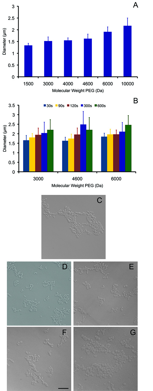
Microgel size is dependent on polymerization conditions. Figure 2A illustrates how microgel diameter increases with increasing PEG starting molecular weight and crosslinking time. Representative images of microgels used for sizing for various molecular weights and crosslinked for 30 sec are shown in Figures 2C-G. For PEG with a molecular weight of 3,000 Da, the microgel average diameter increased from 1.65±0.26 to 2.20±0.54 μm as UV exposure increased from 30-600 sec (Figure 2B). With increasing molecular weight from 3,000-10,000 Da while holding UV exposure constant at 30 sec, microgel diameter increased from 1.65±0.26 to 2.17±0.33 μm (Figure 2A). Significant differences were not observed between molecular weights of 3,000, 4,000, and 4,600 Da. A detailed account of statistically significant differences with the precipitation-formed microgels are in Tables 1 and 2. Longer polymerization time lends to more crosslinking with a greater opportunity for multiple chains to crosslink and precipitate. Additional UV exposure allowed for some microgels to form from chains that had a larger polymer chain length resulting in a more heterogeneous size distribution as indicated by the increased distribution in microgel diameter. Representative histograms are shown for PEG 3,000 in Figures 3A-E and results for all molecular weights in Figure 3F. Considering PEG 3,000 for 30-600 sec of UV exposure, PDI increased from 1.12-2.34, but for shorter crosslinking times (30 sec) PDI remained low for all molecular weights 1.04, 1.14, 1.04, 1.14, 1.12, and 1.21 for PEG 1,500, 3,000, 4,000, 4,600, 6,000, and 10,000 Da, respectively.
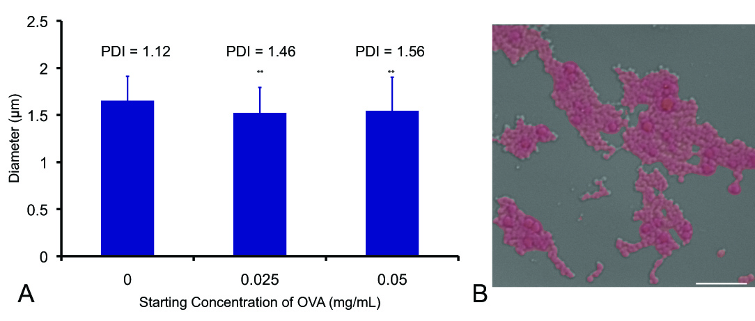
Microgel diameter and PDI were also examined for microgels formed with different concentrations of fluorescently-labeled ovalbumin (OVA-555) in solution (step 2.2) prior to crosslinking. The OVA-555 in solution was encapsulated as the crosslinked PEG chains precipitated and formed microgels, shown in Figure 4B. Microgel diameter was significantly impacted by encapsulated OVA when compared to blank microgels. Additionally, microgels with encapsulated OVA-555 had a greater PDI than those without (Figure 4A). The average diameter was 1.65±0.26 μm for blank microgels, 1.52±0.27 μm with 0.025 mg/ml OVA-555, and 1.54±0.36 μm with 0.05 mg/ml OVA-555. PDI increased from 1.12 for blank microgels to 1.56 for 0.05 mg/ml OVA-555.
The density, a measure of the polymer content, was examined for PEG 3,000, 4,600, and 6,000 at each crosslinking time (Figure 5A). Microgels formed with 20% PEG 3,000 typically resided between solutions of 4-5% dextran, while microgels formed with PEG 4,600 were located between 3-4% dextran, and microgels formed with PEG 6,000 were between 3-1% dextran; these densities were calculated to be 1.016, 1.015, and 1.012 g/cm3 respectively. No statistical differences of microgel density within each MW for crosslinking times of 30-600 sec were observed. Additionally, no differences in density were noted between microgels formed from PEG 3,000 and PEG 4,600, however all crosslinking times for microgels with PEG 6,000 were statistically different from both PEG 3,000 and PEG 4,600. The increased density in the lower molecular weight correlated well with swelling results (Figure 5B). The microgels’ ability to swell in water was similar for all crosslinking times within each MW grouping, with approximately 95, 96, and 96% water imbibed for MW of 3,000, 4,600, and 6,000 respectively. The swelling, which is a measure of the amount of water in the microgels at equilibrium, was significantly different for all crosslinking times between microgels from PEG 3,000 and PEG 6,000.

Figure 1. Reaction schematic for the crosslinking of the microgels. Click here to view larger image.

Figure 2. Chart for average diameter ± standard deviation of PEG microgels with a starting concentration of 20% PEG. (A) MW of 1,500, 3,000, 4,000, 4,600, 6,000, and 10,000 Da and UV exposure of 30 sec. ANOVA was performed with p=0.05 set for significance level. All MWs were significantly different from each other with the exception of PEG 3,000, 4,000, and 4,600 Da. (B) PEG microgels with MW of 3,000, 4,600, and 6,000 Da and UV exposure of 30, 90, 120, 300, and 600 sec. For each MW and crosslinking time, the minimum number of microgels measured was 37. (C-G) Representative images of microgels after 30 sec of crosslinking at the various molecular weights with (C) PEG 1,500 (D) PEG 3,000 (E) PEG 4,600 (F) PEG 6,000 and (G) PEG 10,000. Microgels were imaged using 100X DIC oil objective and are all at the same magnification, scale bar = 10 μm. Click here to view larger image.

Figure 3. Evaluation of microgel size distribution and dispersity. Histogram of diameters for PEG microgels with a starting concentration of 20% PEG, MW 3,000 Da and crosslinking times of 30 (A), 90 (B), 120 (C), 300 (D), and 600 sec (E), bin size =0.2. Variations in polydispersity with changes in starting MW and crosslinking time are also represented (F). For each MW and crosslinking time examined, a minimum of 37 microgels were measured. Click here to view larger image.

Figure 4. OVA conjugated with Alexa Fluor 555 was mixed into the solution prior to UV exposure. Chart for average diameter ± standard deviation for each concentration of OVA-555 in solution prior to crosslinking with PDI noted for each sample group. Significant differences were found between loaded and unloaded microgels (noted by **), however no significant difference were found between 0.025 and 0.05 mg/ml (A). Microgels with encapsulated OVA-555 exhibited fluorescence (B). Control microgels did not exhibit fluorescence confirming there is no PEG auto-fluorescence. (100X oil objective, scale bar=10 μm). Click here to view larger image.

Figure 5. Microgels were characterized via density (A) and swelling (B). These measurements are complementary and characterize the amount of polymer within the microgels. (A) The density was measured via isopynic dextran gradients, and microgels formed with either PEG 3,000 or PEG 4,600 were not statistically different from each other. For each crosslinking time, microgels formed from PEG 6,000 had statistically lower density that either PEG 3,000 or PEG 4,600, denoted by * (p<0.05). (B) Swelling was measured using mass of microgels in water at equilibrium and dry mass. Statistical differences from PEG 3,000 (similar time) are denoted by * (p<0.05) and from PEG 4,600 (similar time) are denoted by # (p<0.05). Click here to view larger image.
Diskussion
Physical properties of PEG microgels were examined for changes in polymerization conditions. For this precipitation reaction, a buffer solution, 20% (w/v) PEG-diacrylate (MW 3,000, 4,600, or 6,000 Da) solution, and photoinitiator were mixed and warmed to 37 °C. Addition of 1.5 M Na2SO4, a kosmotrophic salt that increases the interactions between water molecules, caused PEG to momentarily precipitate upon its addition. This effect is more prominent with higher molecular weight PEG8, suggesting that actual microgel formation is a combination of chain growth and precipitation. Microgels were obtained from photopolymerization with ultraviolet (UV) light. UV exposure was varied from 30, 90, 120, 300, and 600 sec. When the polymer reaches a critical molecular weight it precipitates, forming entangled and crosslinked microgels. Encapsulation of protein into the microgels was accomplished by addition of the molecule to the solution prior to UV exposure. As the polymer chains form crosslinks, they entrap the protein in the polymer network2,4.
Microgels were measured for size and PDI by optical microscopy in conjunction with Axiovision software. A volume-based equation was used to determine PDI, with a PDI of 1 being monodisperse. Increasing the PEG molecular weight increased the microgel size made from PEG 3,000 to PEG 6,000; increasing time to UV exposure increased PDI. Microgels formed from PEG 4,600 did not show any significant differences to PEG 3,000 or PEG 6,000, likely due to overlap in the molecular weight distributions of the reactants. However, a previous study found a significant difference in diameter between PEG 3,000 (1.62±0.07 μm) and PEG 3,400 microgels (2.39±0.05 μm)11, which should have more overlap in molecular weight distribution than PEG 3,000 and 4,600.
Encapsulation of OVA-555 significantly impacted microgel diameter, however this lab has previously shown that surface modifications do not impact microgel diameter13. Entrapping the OVA, which has a radius of gyration of approximately 50 Å14, likely increased the diameter simply by occupying space within the microgels. Similarly, we expect the PDI to increase due to the possible differences in amount of OVA per microgel. Preliminary data (not shown) demonstrated that at lower OVA concentrations, not all microgels encapsulated sufficient OVA to image via fluorescence. The total amount of protein encapsulated would be dependent upon the size of the protein, in addition to other factors such as shape and hydrophobicity. In addition, a maximum encapsulation mass per mass of microgels is likely based on preliminary data, and should be considered when examining encapsulation efficiency. Further analysis on the amount of encapsulation and potential release are necessary to fully characterize the loaded microgels.
While well correlated to each other, the density and swelling measurements resulted in different amounts of polymer in the microgels, likely due to the methods related to the measurement. For example, any equilibrium of the dextran solution with the microgels would result in a measurement of increased polymer content. In contrast, the swelling measurement required removal of all residual water surrounding the microgels. If excess water was imbibed within the system, the calculated results would overestimate the amount of water. Therefore, it was important to have several methods to quantify the amount of polymer in the system.
These multiple measurements are particularly important for the microgel system. While bulk hydrogels incorporate most of the polymer added due to the method of casting, not all of the 20% PEG initially added was incorporated into the microgels. Comparing directly to bulk, PEGDA hydrogels, the water content reported for 5% PEG hydrogels previously15 was comparable to the water content in PEG microgels formed with 20% PEG 3000 (95%). In contrast, 4 kDa 20% PEG hydrogels had a water content of 83±2%9, while PEG 4600 microgels in this study had a water content of ~96%. Even with different initiators and precursors, similar trends were observed by a variety of bulk-crosslinked PEG hydrogels15-19. In addition, increasing the starting PEG MW decreased microgel density from 1.016, 1.015, and 1.012 g/cm3 with PEG 3,000, 4,600, and 6,000, respectively. Microgels formed from a higher MW resulted in less tightly knit microgels with greater ability to swell and, therefore, a lower density. Again, this trend has been noted in the literature with bulk-PEG hydrogels19. The results from this study, as well as results from the literature20-22 indicate that altering polymer MW will be an effective method of modifying microgel properties to influence the release of solutes.
These changes in starting MW could not overcome one limitation of the precipitation reaction — the narrow range of diameters produced. Using PEG 1,500-10,000, microgel diameter ranged from 1.34±0.09 to 2.17±0.33 μm. Additionally, preliminary observations have shown little effect on microgel size when modifying the starting Na2SO4 concentration and starting PEGDA concentration in the precursor. Increased salt concentration initiated faster precipitation while increased PEGDA concentration increased microgel diameter slightly and decreased water content in the microgels (data not shown).The swollen size of the PEG microgels was correlated to the molecular weight of the reactants, similar to published reports on higher molecular weights PEG form hydrogels that had larger mesh size and lower elasticity3,9. In contrast to the precipitation-reaction, molecular weight of the starting PEG did not significantly impact the diameter of microspheres produced by emulsification23. Additionally, if nanoparticles are desired, emulsion polymerization may be the preferred technique as this typically produces particles on the nano-scale from high molecular weight polymers24. Suspension polymerization, on the other hand, is the preferred method for larger particles 10 μm-5 mm25. However, the nature of emulsion reactions yield highly disperse particles23, which would alter the release kinetics if utilized for drug delivery26. In addition to the issues with polydispersity, the other formation techniques require the use of solvents. Toxicity issues due to solvents have been reduced by the use of acetone as the dispersed phase because acetone has a low toxic potential to humans27. However, solvent contact may significantly reduce the bioactivity and bioavailability of encapsulated molecules, limiting the range of molecules that can be encapsulated.
The nature of the precipitation reaction offers several benefits over existing systems. Reaction conditions are mild and organic solvents and surfactants are not required to form or stabilize the microgels. Additionally, the reaction produces spherical microgels with a low PDI, which show great potential for controlled release vehicles.
Offenlegungen
The authors declare that they have no competing financial interests.
Danksagungen
Funding for this project was through NSF CBET Award 1061834. The authors would like to acknowledge CIBA for a sample of photoinitiator.
Materialien
| Name | Company | Catalog Number | Comments |
| Phosphate Buffered Saline (PBS) | MP Biomedical | 2810305 | |
| Triethanolamine (TEOA) | J.T. Baker | 9468-01 | Preheat to 37 °C prior to pipetting |
| Hydrochloric acid (HCl) | BDH Aristar | BDH3028 | |
| Sodium Sulfate | J.T. Baker | 3891-01 | |
| Irgacure 2959 | Ciba | 029891301PS04 | |
| Ovalbumin (OVA) | Invitrogen | 34782 | |
| PEG 1,500 | Alfa Aesar | A16241 | |
| PEG 3,000 | Fluka | 03997-1KG | |
| PEG 4,000 | Alfa Aesar | A16151 | |
| PEG 4,600 | Sigma | 373001-250G | |
| PEG 6,000 | Fluka | 03394-1KG | |
| PEG 10,000 | Alfa Aesar | B21955 | |
| Dextran 70 | TCI | D1449 |
Referenzen
- McNaught, A. D., Wilkinson, A. . IUPAC Compendium of Chemical Terminology (The Gold Book). 2nd edn. , (1997).
- Lu, S. X., Anseth, K. S. Release behavior of high molecular weight solutes from poly(ethylene glycol)-based degradable networks. Macromolecules. 33, 2509-2515 (2000).
- Peppas, N. A. . Hydrogels in Medicine. 1, (1986).
- West, J. L., Hubbell, J. A. Photopolymerized hydrogel materials for drug delivery applications. React. Polym. 25, 139-147 (1995).
- Hunkeler, D., et al. . Theories and Mechanism of Phase Transitions, Heterophase Polymerizations, Homopolymerization, Addition Polymerization. Advances in Polymer Science. 112, 115-133 (1994).
- Arshady, R. Suspension, emulsion, and dispersion polymerization: A methodological survey. Colloid Polym. Sci. 270, 717-732 (1992).
- Bai, F., Yang, X. L., Huang, W. Q. Synthesis of narrow or monodisperse poly(divinylbenzene) microspheres by distillation-precipitation polymerization. Macromolecules. 37, 9746-9752 (2004).
- Bailey, F., Callard, R. W. Some properties of poly(ethylene oxide) in aqueous solution. J. Appl. Polym. Sci. 1, 56-62 (1959).
- Cruise, G. M., Scharp, D. S., Hubbell, J. A. Characterization of permeability and network structure of interfacially photopolymerized poly(ethylene glycol) diacrylate hydrogels. Biomaterials. 19, 1287-1294 (1998).
- Flory, P. . Principles in Polymer Chemistry. , (1953).
- Flake, M. M., et al. Poly (ethylene glycol) microparticles produced by precipitation polymerization in aqueous solution. Biomacromolecules. 12, 844-850 (2011).
- Sawhney, A. S., Pathak, C. P., Hubbell, J. A. Bioerodible hydrogels based on photopolymerized poly(ethylene glycol)-co-poly(.alpha.-hydroxy acid) diacrylate macromers. Macromolecules. 26, 581-587 (1993).
- Scott, R. A., Elbert, D. L., Willits, R. K. Modular poly(ethylene glycol) scaffolds provide the ability to decouple the effects of stiffness and protein concentration on PC12 cells. Acta Biomaterialia. 7, 3841-3849 (2011).
- Ianeselli, L., et al. Protein-Protein Interactions in Ovalbumin Solutions Studied by Small-Angle Scattering: Effect of Ionic Strength and the Chemical Nature of Cations. J. Phys. Chem. B. 114, 3776-3783 (2010).
- Scott, R., Marquardt, L., Willits, R. K. Characterization of poly(ethylene glycol) gels with added collagen for neural tissue engineering. J. Biomed. Mater. Res. A.. 93, 817-823 (2010).
- Lin, H., Kai, T., Freeman, B. D., Kalakkunnath, S., Kalika, D. S. The Effect of Cross-Linking on Gas Permeability in Cross-Linked Poly(Ethylene Glycol Diacrylate). Macromolecules. 38, 8381-8393 (2005).
- Mellott, M. B., Searcy, K., Pishko, M. V. Release of protein from highly cross-;linked hydrogels of poly(ethylene glycol) diacrylate fabricated by UV polymerization. Biomaterials. 22, 929-941 (2001).
- Bryant, S. J., Anseth, K. S., Lee, D. A., Bader, D. L. Crosslinking density influences the morphology of chondrocytes photoencapsulated in PEG hydrogels during the application of compressive strain. J. Orthop. Res. 22, 1143-1149 (2004).
- Padmavathi, N. C., Chatterji, P. R. Structural Characteristics and Swelling Behavior of Poly(ethylene glycol) Diacrylate Hydrogels. Macromolecules. 29, 1976-1979 (1996).
- Ross, A. E., Tang, M. Y., Gemeinhart, R. A. Effects of molecular weight and loading on matrix metalloproteinase-2 mediated release from poly(ethylene glycol) diacrylate hydrogels. AAPS J. 14, 482-490 (2012).
- Sun, G., Zhang, X. -. Z., Chu, C. -. C. Effect of the molecular weight of polyethylene glycol (PEG) on the properties of chitosan-PEG-poly(N-isopropylacrylamide) hydrogels. J. Materi. Sci. Mater. Med. 19, 2865-2872 (2008).
- Zustiak, S. P., Leach, J. B. Characterization of Protein Release From Hydrolytically Degradable Poly(Ethylene Glycol) Hydrogels. Biotechnol. Bioeng. 108, 197-206 (2011).
- Ruan, G., Feng, S. S. Preparation and characterization of poly(lactic acid)-poly(ethylene glycol)-poly(lactic acid) (PLA-PEG-PLA) microspheres for controlled release of paclitaxel. Biomaterials. 24, 5037-5044 (2003).
- Loxley, A., Vincent, B. Equilibrium and kinetic aspects of the pH-dependent swelling of poly(2-vinylpyridine-co-styrene) microgels. Colloid Polym. Sci. 275, 1108-1114 (1997).
- Vivaldo-Lima, E., Wood, P. E., Hamielec, A. E., Penlidis, A. An updated review on suspension polymerization. Ind. Eng. Chem. Res. 36, 939-965 (1997).
- Ritger, P. L., Peppas, N. A. A simple equation for description of solute release I. Fickian and non-fickian release from non-swellable devices in the form of slabs, spheres, cylinders or discs. Journal of Controlled Release. 5, 23-36 (1987).
- Matsumoto, A., Kitazawa, T., Murata, J., Horikiri, Y., Yamahara, H. A novel preparation method for PLGA microspheres using non-balogenated solvents. J. Controlled Release. 129, 223-227 (2008).
Nachdrucke und Genehmigungen
Genehmigung beantragen, um den Text oder die Abbildungen dieses JoVE-Artikels zu verwenden
Genehmigung beantragenThis article has been published
Video Coming Soon
Copyright © 2025 MyJoVE Corporation. Alle Rechte vorbehalten