Method Article
Comparison of Three Clinical Stereoscopic Methods for Measuring Binocular Visual Function During Amblyopic Treatment in Unilateral Amblyopia
In This Article
Summary
Here, we present three common stereopsis measurement methods to measure patients with amblyopia and the comparison of the efficacy of different methods.
Abstract
This study aimed to compare the measuring stereopsis results of unilateral amblyopia during amblyopic treatment by employing some of the most widely used clinical tests. Thirty-four individuals with previously untreated unilateral amblyopia, aged 8.4 ± 2.7 years, were included in the study. Monocular (best corrected visual acuity [BCVA] at distance) and binocular (including Titmus, Random-dot, and Frisby stereopsis) visual functions were measured at baseline and 2-month and 6-month visits after synthetical treatment.
We found that Titmus stereopsis was always significantly better than Random-dot stereopsis (p < 0.001). Frisby stereopsis was also always significantly better than Random-dot stereopsis (p < 0.001). However, there was no significant difference between Titmus stereopsis and Frisby stereopsis (p = 0.562). However, interestingly, there was no significant difference in the mean improvement of the three stereopsis methods from baseline to the 2-month visit, F = 1.158, p = 0.318.
Similarly, a significant difference was also lacking in the mean improvement of the three stereopsis methods from baseline to the 6-month visit, F = 0.302, p = 0.740. We conclude that there will be different results obtained from different stereopsis measuring methods used to measure amblyopia in patients. We recommend performing at least two types of stereoscopic measurements to evaluate each case of amblyopia. However, for observing therapeutic effects, each measurement method has the same performance for clinical results during amblyopic treatment.
Introduction
Amblyopia is a disorder of development of the visual system that results from abnormal visual input during the critical phase of early development, affecting between 1% and 5% of the general population1,2,3,4. It often affects only one eye, caused by strabismus, anisometropia, or form deprivation during visual development. Persons with unilateral amblyopia are clear-sighted under normal everyday viewing conditions, but the vision is dominated by the strong eye. Webber suggested that reduced stereopsis is the most common visual deficit associated with amblyopia5. Furthermore, stereopsis is more degraded by monocular blur (or monocular contrast reduction) than by both eyes being blurred6,7. Even if the visual acuity (VA) of the amblyopic eye has been successfully corrected, their stereopsis remained impaired8.
Impairment associated with amblyopia may have a great influence on persons' lives: visually guided hand movements take longer and are less accurate under monocular viewing conditions than under binocular vision9. Moreover, the performance of motor skills tasks is related to the subject's stereoacuity, and in a large cohort of children and adults, those with normal stereoacuity were found to perform motor tasks best10. Walking performance is also significantly degraded; relative to people with normal binocular viewing, walkers are slowed by ~10% in monocular vision conditions and raise their feet higher when stepping over obstacles11. It has also been reported that reduced stereoacuity affects more complex visuomotor tasks, including reading ability in children aged 5-6 years and academic performance in reading, writing, mathematics, and spelling ability in 5- to 9-year-olds12,13. Impaired stereopsis may also limit career options for amblyopes.
Therefore, in the past few decades, we have come to realize that restoration of VA is not the only goal of amblyopia management. Rather, the recovery of a patient's binocular visual function, especially in terms of stereopsis, should be paid attention to. Previous research has demonstrated that binocular function can be improved with the enhancement of VA in the process of amblyopia treatment14,15,16. For example, Lee showed that when employing occlusion therapy for amblyopia, as VA improves, stereopsis generally improves alongside it (using the Titmus test)14. Wallace found that better outcome related to stereoacuity (using the Randot Preschool Stereoacuity test) was associated with better baseline stereoacuity and better amblyopic eye acuity as an outcome15. Stewart found that stereoacuity (using the Frisby test) improved for almost one-half of the study participants after treatment16. However, the methods used for measuring and quantifying stereopsis in their study were significantly different. Therefore, the differences between these improvements cannot be compared.
The stereoacuity results are also affected by the measurement method. For example, in the widely used Randot 'Circles' Test, the circles are presented on a background of random dots. However, these dots are highly visible monocularly, which may be conducive to amblyopic patients with poor vision still seeing them17. Another widely used stereoscopic test is the Titmus Stereo Test. However, it also has monocular clues, which give rise to false positive results18. For instance, the inclusion of monocular cues makes it possible for subjects to pass the initial two to four levels of the test without stereopsis18,19. Similarly, in the Frisby test, it was found that several children with amblyopia who passed the test had interocular differences of 4-5 lines in VA20. Another point is that the repeatability and agreement in stereoacuity measures are different. The repeatability of stereoacuity measures has been found to be low in patients with poor binocular vision but fairly good in patients with normal binocular vision except on the Random-dot stereopsis test21.
As mentioned above, stereopsis is a binocular visual function that is particularly important to everyone. However, there are many issues hindering the measurement of stereopsis in reality (e.g., limitations of the measurement method, repeatability, agreement). Previous research mainly focused on one stereopsis test as the standard for recovery. Therefore, we ask the following question: Is this standard reliable? If we use different stereopsis measurement methods to evaluate the recovery of stereopsis, will the results be the same or different? When there is a change during the treatment for amblyopia using a given stereopsis measurement, how do we know whether the change is sufficient to be clinically significant or whether the results may be attributed to the technique used for measurement? To address these questions, in the present study, we compared the stereopsis measurement results for unilateral amblyopia during amblyopia treatment using three following clinical tests, which are some of the most widely used approaches: Random-dot stereopsis, Titmus stereopsis, and Frisby stereopsis. The study aimed to evaluate the differences in the three stereopsis measurement methods related to measuring the degree of improvement of binocular visual function during the treatment of amblyopia and to find the correlations between them.
Protocol
The study protocol was approved by the institutional review board of Anhui Medical University. Written informed consent was obtained from each participant's parent or legal guardian after an explanation of the nature and possible consequences of the study. The methods and data collection were carried out under approved guidelines. Thirty-four individuals with unilateral amblyopia, including 21 boys and 13 girls aged 8.4 ± 2.7 years, were included in this study. They were recruited from the Department of Ophthalmology of the Second Affiliated Hospital of Anhui Medical University (Anhui, China).
1. Patient selection
- Select patients who have been newly diagnosed with unilateral amblyopia and have never received any treatment (including spectacle wear or occlusion) before the study.
- Perform a complete initial ophthalmological examination on all the participants, including cycloplegic refraction, best corrected visual acuity (BCVA) at distance (5 m) for each eye, eye alignment by cover testing, slit-lamp biomicroscopy for anterior segment examination, and dilated fundus examination.
- Set the inclusion criteria as unilateral amblyopia, including anisometropic amblyopia and strabismic amblyopia.
- Set the exclusion criteria of this study to include a history of prior treatment with spectacles, a history of occlusion or penalization therapy, corneal opacity, cataract, glaucoma, ocular pathology, and previous eye surgery.
- After performing objective streak retinoscopy, prescribe spectacles to all patients (except those with strabismic amblyopia) based on cycloplegic refraction with the following prescriptions:
- Fully correct myopic and astigmatic refractive errors.
- Correct hyperopic refractive errors within 1.00 D of full correction.
- Correct anisometropia to less than 1.00 D.
NOTE: Since the improvement in stereopsis during amblyopia treatment is self-comparison, and the purpose is to compare the differences between the three stereoscopic measurements, do not allow patients with anisometropic amblyopia to undergo refractive adaptation.
- Perform synthetical treatment for patients who need refractive correction. Prescribe continuous4 h of patching per day to all participants and ask the patients who need refractive correction to wear spectacles all day long. At the follow-up visit, ask the patients and/or their parents how long the spectacles were worn and how long the occlusion was performed each day. Include only patients with good reported compliance in the study.
2. Visual function test
- Measure monocular (BCVA at distance) and binocular (including Titmus stereopsis, Random-dot stereopsis, and Frisby stereopsis) visual function at baseline and 2 months and 6 months after treatment. Ensure that all the VA tests are conducted by the same nurse, who must be uninformed as to the purpose of the study, and that all the stereopsis measures are conducted by an ophthalmologist.
- Evaluate stereopsis in a silent room with sufficient light (500 lux) using the Titmus (butterfly, animals, and rings), Random-dot (animals and forms tests), and Frisby stereopsis. Before the measurement begins, carry out proper demonstrations of the test and give enough resting time to the participants to avoid fatigue and its negative effects on the test results.
- Carry out the three stereopsis measurement methods randomly in each participant and at each measurement; show each optotype line separately. For the statistical analysis, assign a value of 3,000 arcsec to patients who have no stereopsis.
- Best corrected visual acuity (BCVA)
- Prepare the vision chart: measure the participants' best spectacle-corrected VA monocularly using the full Tumbling E Chart at 5 m. Ensure the chart is placed in a well-lit environment.
- Set up the testing environment: conduct the test at a distance of 5 m from the vision chart. If space is limited, use mirrors to create an equivalent 5 m visual path.
- Explain the test process: Explain to the patient the purpose and steps of the BCVA test. Ensure that the patient understands that the test aims to determine how well they can see under optimal vision correction.
- Ensure that the patient wears corrective lenses or glasses during the test if they normally wear them.
- Test each eye separately: examine the amblyopic eye first during the experiment. Cover one eye and test the vision of the other, then switch and test the other eye. Make sure the patient's eyes do not become fatigued from prolonged focusing.
- Record vision readings: ask patients to read the optotypes one after another and stop them when they cannot respond within 10 s by using a stopwatch. Note the smallest line of vision that the patient can clearly identify.
- Evaluate the results: assess the patient's best corrected visual acuity based on the recorded vision values. Express all VA scores as the logarithm of the minimum angle of resolution (logMAR) for the analyses.
- Titmus stereopsis
- Prepare the equipment (see the Table of Materials): ensure that the Titmus stereoscopic vision tester and its accompanying stereoscopic test cards are in good condition. Perform Titmus stereopsis at a 40 cm distance with polarized glasses over the best-refractive-corrected glasses.
- Setting the environment: choose a room with appropriate lighting for the test, avoiding strong direct sunlight or overly dim conditions.
- Explain to the patient the purpose and process of the test. Make sure the patient understands that the images they will see might appear to "jump out" from the surface of the card. Give enough resting time to the participants to avoid fatigue and its negative effects on the test results.
- Have the patient wear the special polarized glasses. The glasses help the patient to correctly perceive the stereoscopic images on the test cards.
- Conducting the test:
- Conduct the Butterfly test first. Ask the patient to identify different parts of the stereoscopic butterfly, such as wings, antennae, etc.
- Conduct the Circle test and ask the patient to observe a series of circles and determine which circle appears to "stand out" more than the others.
- Conduct the Animal test for children, involving the identification of different animal images.
- Record the patient's performance on different test cards, paying special attention to their ability to identify stereoscopic images.
- Have the patient remove the stereoscopic glasses after completing the examination.
- Assess the stereoscopic vision capabilities of the patients based on their responses. Poor stereoscopic vision may indicate depth perception issues or other visual impairments.
NOTE: During the Titmus stereoscopic vision test, ensure the patient is comfortable throughout the process and adjust your explanations according to their age and level of understanding.
- The Random-dot stereopsis
- Prepare the test materials: get the Random-dot stereopsis test charts ready. These charts typically consist of seemingly random dots that form a stereoscopic image when viewed with special glasses. The Random-dot stereopsis consists of seven Random-dot stereograms, which can be seen in depth perception through red and green filters.
- Set up the testing environment: choose a room with even lighting for the test, avoiding strong direct sunlight or overly dim conditions.
- Explain to the patient the purpose and steps of the test. Inform them that they will be trying to identify hidden images within what appears to be a random dot pattern. Give enough resting time to the participants to avoid fatigue and its negative effects on the test results.
- Have the patient wear the red and green filters, which will help the patient differentiate the various layers in the test chart, creating a stereoscopic effect.
- Conduct the test; present the Random-dot test chart to the patient wearing the stereoscopic glasses. Ask them to describe the images they see. This may include shapes, patterns, or specific figures. Participants identify the shapes or figures through the red and green filters over their best-refractive-corrected glasses at a distance of 40 cm.
- Note the patient's ability to recognize the stereoscopic images. Focus on whether they can accurately identify the stereoscopic images and the time it takes to do so. The results range disparities from 800 to 40 arcsec.
NOTE: In this study, participants were classed as 'negative' responders if they were unable to correctly identify the largest shape and allocated a notional score of 3,000 arcsec. - Have the patient remove the filters after completing the examination.
- Assess the stereoscopic vision of the patients based on their ability to recognize stereoscopic images. Difficulty in identifying or misidentifying images may indicate a problem with stereoscopic vision functions. When conducting the Random-dot stereopsis test, ensure that the patient is comfortable throughout the process and adjust explanations according to their age and level of understanding. If necessary, repeat certain parts of the test to ensure the accuracy of the results.
- Frisby stereopsis
- Prepare the test materials: the Frisby stereopsis test consists of several transparent plastic plates of different thicknesses, each embedded with a specific pattern. As the Frisby stereopsis is a very real test since the background and target are seen in depth, special glasses are not required. The test equipment consists of transparent plates, each subdivided into four squares with different-sized and randomly placed arrowheads printed onto one surface and with one square containing a circular target of arrowheads printed onto the other surface.
- Set up the testing environment: choose a well-lit, distraction-free environment for the test. Ensure that the testing area has uniform lighting, avoiding strong direct sunlight or overly dim conditions.
- Explain to the patient the purpose and basic steps of the Frisby stereopsis test. Ensure the patient understands that the test aims to assess their stereoscopic vision capabilities. Give enough resting time to the participants to avoid fatigue and its negative effects on the test results.
- As pretest preparation, ask the patient to sit approximately 40 cm away from the test plates. If patients require glasses, ask them to wear them during the test.
- For conducting the test, use the test plates of different thicknesses in succession. Each time, place a plate in front of the patient and ask if they can see the pattern in the plate "pop out."
- Note the patient's responses to each plate, paying special attention to whether they can correctly perceive the stereoscopic effect of the pattern.
- Assess their stereoscopic vision based on their responses to plates of different thicknesses. Thinner plates require stronger stereoscopic vision capabilities to recognize the pattern. The test has three plates of different thicknesses-namely, 6 mm, 3 mm, and 1.5 mm-with respective values of 340 arcsec, 170 arcsec, and 55 arcsec at a distance of 40 cm.
NOTE: A wide range of disparities can be obtained using sheets of different thicknesses. - Analyze the results and determine the level of their stereoscopic vision based on their responses to the test plates. When conducting the Frisby stereopsis test, ensure the patient is comfortable throughout the process and adjust explanations according to their age and level of understanding. If necessary, repeat certain steps of the test to ensure the accuracy of the results.
NOTE: Inability to correctly perceive the stereoscopic effect on all plates may indicate a stereoscopic vision disorder.
3. Statistical methods
- For statistical analysis, convert VA to logMAR.
- Assign participants who have no stereopsis a stereoacuity of 3,000 arcsec. Convert stereoacuity scores in seconds of arc to log values.
- Compare VA or the levels of stereopsis before and after the treatment using paired-samples t-tests.
- Use repeated measures analysis of covariance to compare the differences of the three stereopsis measures at different visits. Use an analysis of covariance to compare the differences in the mean improvement of the three stereopsis methods at different visits.
- Consider p-values < 0.05 significant.
Results
Visual benefits of synthetic treatment in unilateral amblyopia
BCVA
The mean amblyopic eye BCVA improvement from baseline to the 2-month visit with synthetical treatment was 0.19 ± 0.14 logMAR (95% confidence interval [CI] of the difference was [0.14, 0.24]), and this improvement was statistically significant (mean BCVA ± standard deviation [SD] at baseline, 0.60 ± 0.24 vs 2-month visit, 0.41 ± 0.21, t (33) = 7.903, p < 0.001; Figure 1A). At the 6-month visit, the improvement from baseline was 0.30 ± 0.15 logMAR (95% CI of the difference was [0.24, 0.35]), and this improvement was also significant (mean BCVA ± SD at the 6-month visit, 0.31 ± 0.19, t (33) = 11.547, p < 0.001; Figure 1B).
Random-dot stereopsis
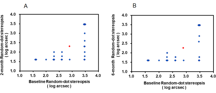
The mean Random-dot stereopsis of the participants significantly improved from baseline to the 2-month visit with synthetical treatment (mean ± SD at baseline, 2.89 ± 0.70 arcsec vs 2-month visit, 2.32 ± 0.82 arcsec; t (33) = 5.501, p < 0.001; Figure 2A). This improvement was 0.57 ± 0.60 arcsec, and 95% CI of the difference was [0.36, 0.78]. At the 6-month visit, the improvement from baseline was 0.62 ± 0.70 (95% CI of the difference was [0.38, 0.85]), and this improvement was also significant (mean stereopsis ± SD at the 6-month visit, 2.27 ± 0.84, t (33) = 5.283, p < 0.001; Figure 2B).
Titmus stereopsis
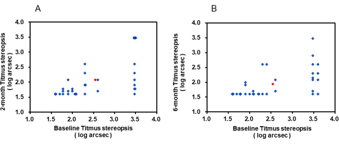
The mean Titmus stereopsis of the participants at baseline was 2.54 ± 0.75 arcsec before the treatment, and it significantly improved to 2.08 ± 0.65 arcsec at the 2-month visit, t (33) = 4.465, p < 0.001 (Figure 3A). The mean improvement from baseline to the 2-month visit with synthetical treatment was 0.46 ± 0.60 arcsec (95% CI of the difference was [0.25, 0.67]). At the 6-month visit, the improvement from baseline was 0.60 ± 0.61 arcsec (95% CI of the difference was [0.38, 0.81]), and this improvement was also significant (mean stereopsis ± SD at the 6-month visit, 1.94 ± 0.48, t (33) = 5.677, p < 0.001; Figure 3B).
Frisby stereopsis
The mean Frisby stereopsis of the participants at baseline was 2.39 ± 0.71 arcsec before the treatment, and it significantly improved to 2.05 ± 0.58 arcsec at the 2-month visit, t (33) = 3.222, p = 0.003; (Figure 4A). The mean improvement from baseline to the 2-month visit with synthetical treatment was 0.35 ± 0.63 arcsec (95% CI of the difference was [0.13, 0.56]). At the 6-month visit, the improvement from baseline was 0.50 ± 0.59 arcsec (95% CI of the difference was [0.30, 0.71]), and this improvement was also significant (mean stereopsis ± SD at the 6-month visit, 1.89 ± 0.37, t (33) = 4.952, p < 0.001; Figure 4B).
Comparison of the three clinical stereopsis tests for unilateral amblyopia during synthetical treatment
The method of repeated measurement analysis of variance was used to analyze whether stereopsis measured by the three methods were different with the change in time. The effect of the interaction between measurement method and time on the measurement method was not statistically significant, F (2.377, 78.438) = 0.934, p = 0.411. Therefore, it is necessary to interpret the main effects of the two subjects' internal factors (measurement method and time).
The time factor had statistical significance to the stereopsis, F (1.602, 52.855) = 43.843, p < 0.001. The stereopsis at baseline was 0.458 arcsec higher than that of stereopsis at the 2-month visit (95% CI: 0.269-0.648), and the difference was statistically significant (p < 0.001). The stereopsis at baseline was 0.572 arcsec higher than that of stereopsis at the 6-month visit (95% CI: 0.399-0.746), the difference was also statistically significant (p < 0.001). The stereopsis at the 2-month visit was 0.114 higher than that of stereopsis at the 6-month visit (95% CI: -0.003-0.231) arcsec, but the difference was not statistically significant (p = 0.059).
The main effect of the measurement method on stereopsis was statistically significant, F (2,66) = 19.553, p < 0.001. The difference between Random-dot stereopsis and Titmus stereopsis was statistically significant (p < 0.001), Titmus stereopsis was significantly better compared with Random-dot stereopsis, the difference was 0.306 (95% CI: 0.154-0.458) arcsec, and the difference between Random-dot stereopsis and Frisby stereopsis was also statistically significant (p < 0.001), Frisby stereopsis was significantly better compared with Random-dot stereopsis, the difference was 0.380 (95% CI: 0.188-0.572) arcsec. However, there was no significant difference between Titmus stereopsis and Frisby stereopsis (p = 0.562), and the difference was 0.074 (95% CI: -0.065-0.213) arcsec. The average stereopsis measured by the three measurement methods is shown in Figure 5.
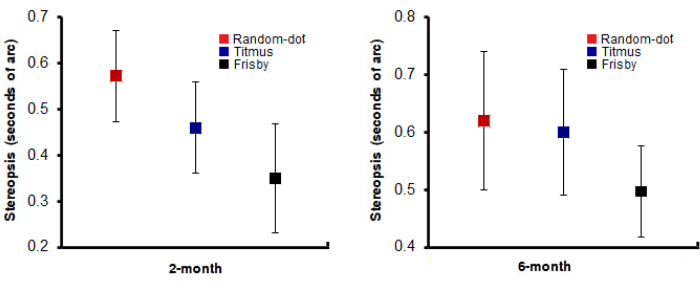
However, interestingly, there was no significant difference in the mean improvement of the three stereopsis methods from baseline to the 2-month visit, F = 1.158, p = 0.318. Furthermore, there was also no significant difference between the mean improvement of Random-dot stereopsis and Titmus stereopsis (mean improvement of stereopsis ± SD, -0.57 ± 0.60 arcsec vs -0.46 ± 0.60 arcsec; p = 1.000), or Random-dot stereopsis and Frisby stereopsis (mean improvement of stereopsis ± SD, -0.57 ± 0.60 arcsec vs -0.35 ± 0.63 arcsec; p = 0.394), or Titmus stereopsis and Frisby stereopsis (mean improvement of stereopsis ± SD, -0.46 ± 0.60 arcsec vs -0.35 ± 0.63 arcsec; p = 1.000) from baseline to the 2-month visit by Bonferroni test.
Similarly, a statistically significant difference was also lacking in the mean improvement of the three stereopsis methods from baseline to the 6-month visit, F = 0.302, p = 0.740. Furthermore, there was also no significant difference between the mean improvement of Random-dot stereopsis and Titmus stereopsis (mean improvement of stereopsis ± SD, -0.62 ± 0.68 arcsec vs -0.60 ± 0.61 arcsec; p = 1.000), or Random-dot stereopsis and Frisby stereopsis (mean improvement of stereopsis ± SD, -0.62 ± 0.68 arcsec vs -0.50 ± 0.59 arcsec; p = 1.000), or Titmus stereopsis and Frisby stereopsis (mean improvement of stereopsis ± SD, -0.60 ± 0.61 arcsec vs -0.50 ± 0.59 arcsec; p = 1.000) from baseline to the 6-month visit by Bonferroni test. The average improvements of stereopsis measured by the three measurement methods are shown in Figure 6.

Figure 1: The change in BCVA after 2 and 6 months of synthetical treatment. Scatterplots showing BCVA at baseline versus at the (A) 2-month and (B) 6-month visits for individual participants undergoing synthetical treatment. Thirty-four patients participated in this study. Each dot represents the results of one participant; the red symbol represents their averaged results. Abbreviations: BCVA = best corrected visual acuity; logMAR = logarithm of the minimum angle of resolution. Please click here to view a larger version of this figure.

Figure 2: The change in Random-dot stereopsis (log arcsec) after 2 and 6 months of synthetical treatment. Scatterplots showing Random-dot stereopsis at baseline versus at the (A) 2-month and (B) 6-month visits for individual participants undergoing synthetical treatment. Thirty-four patients participated in this study. Each dot represents the results of one participant; the red symbol represents their averaged results. Please click here to view a larger version of this figure.

Figure 3: The change in Titmus stereopsis (log arcsec) after 2 and 6 months of synthetical treatment. Scatterplots showing Titmus stereopsis at baseline versus at the (A) 2-month and (B) 6-month visits for individual participants undergoing synthetical treatment. Thirty-four patients participated in this study. Each dot represents the results of one participant; the red symbol represents their averaged results. Please click here to view a larger version of this figure.

Figure 4: The change in Frisby stereopsis (log arcsec) after 2 and 6 months of synthetical treatment. Scatterplots showing Frisby stereopsis at baseline versus at the (A) 2-month and (B) 6-month visits for individual participants undergoing synthetical treatment. Thirty-four patients participated in this study. Each dot represents the results of one participant; the red symbol represents their averaged results. Please click here to view a larger version of this figure.

Figure 5: Bar graph showing the average of the three stereopsis (Random-dot, Titmus, Frisby stereopsis) at baseline and 2-month and 6-month visits. Error bars represent standard errors across participants. Please click here to view a larger version of this figure.

Figure 6: Scatterplot showing the mean improvement of the three stereopsis (Random-dot, Titmus, Frisby stereopsis) from baseline to the 2-month and 6-month visits. Error bars represent standard errors across participants. Please click here to view a larger version of this figure.
Table 1: Clinical characteristics of patients with amblyopia. Please click here to download this Table.
Discussion
Study design and participants
The 34 participants with unilateral amblyopia, including 21 boys and 13 girls aged 8.4 ± 2.7 years, were newly diagnosed and had never received any treatment (including spectacle wear or occlusion) before they participated in the study. In the 34 participants, anisometropic amblyopia was most prevalent (25/34, 74%), whereas strabismic amblyopia was least prevalent (9/34, 26%). The clinical details are shown in Table 1.
It is generally thought that worse stereopsis is associated with worse VA of the amblyopic eye in patients with unilateral amblyopia. Previous works on the functional consequences of the loss of stereopsis in patients with amblyopia have shown that stereopsis is functionally important for vision-related quality of life22. However, many studies have demonstrated that stereopsis results would be affected by the measurement method (even the widely used Randot 'Circles' test has monocular clues that will give rise to false positive results)17,18,19,20 or other measurement problems21,23,24. In this study, we compared the differences in the measuring stereopsis results of unilateral amblyopia during the amblyopic treatment using Titmus, Random-dot, and Frisby stereopsis. We found that the three stereopsis measurements were different during the amblyopic treatment, but there were no differences between the improvements of the stereopsis by the three measurement methods at the follow-up visits during the amblyopic treatment.
It is known that stereopsis and VA can be effectively recovered by glasses alone or occlusion treatment15,16,25,26. Lee and Isenberg found that there was a significant linear relationship between VA and stereoacuity14. When employing occlusion therapy for amblyopia, as VA improves, stereopsis (using the Titmus test) generally also improves (the mean stereoacuity improved from 1,167.4 arcsec to 101 arcsec; p < 0.0001). In the present study, we prescribed 4 h of patching per day in combination with spectacles all day long among anisometropic amblyopia patients. We found that all the stereopsis measured by the three methods could be significantly improved. The mean Titmus stereopsis of our patients improved from 1,146 arcsec to 215 arcsec (p < 0.0001), which is similar to Lee's finding. However, this delicate difference is probably owing to the age of the patients (Lee vs this study, mean age: 5.1 years vs 8.4 years) or the period of the treatment (Lee vs this study, mean duration: 36 weeks vs 6 months). The Random-dot and Frisby stereopsis tests had the same results. This suggests that any of the Titmus, Random-dot, and Frisby stereopsis tests can be recommended as an effective method to observe the clinical efficacy of amblyopic treatment.
Were there any differences in the results of these three stereoscopic methods? Which measurement method was more effective and reliable during measurement? To answer these questions, we measured the stereopsis of the patients at each clinic visit by using the three stereopsis methods. We found that the results of the three stereopsis measurements differed during the amblyopic treatment. The Titmus stereopsis or Frisby stereopsis was always better than Random-dot stereopsis during the amblyopic treatment, but there was no significant difference between Titmus and Frisby stereopsis testing. These results suggest that there are differences in stereopsis results when measured with different tests. If we measure the stereopsis of patients with amblyopia using the Titmus test, the actual results may not be as good as the measurements. However, our study included a relatively small number of patients. What is more, although these different results using the three stereopsis methods were the same at the second and third follow-up visits as at the initial visit, the results may be affected by the synchronous amblyopic treatment. In addition, the stereopsis measurement difference may be related to the level of the stereopsis at the initial visit.
For example, Farvardin et al. compared VA testing with the ability of the TNO, Titmus, and Randot stereo tests for the detection of amblyopia27. In their study, mean stereopsis values were 420 ± 297.3 arcsec with the Titmus test and 388 ± 197.2 arcsec with the Randot test, exhibiting opposing results to ours. This contrary result is probably due to the different levels of stereopsis of the participants (Titmus: 1,146 ± 1393 arcsec vs 420 ± 297.3 arcsec; Random-dot stereopsis: 1,702 ± 1404 arcsec vs 388 ± 197.2 arcsec). Although the level of measurement of stereopsis in the third follow-up visit was similar to their results (Titmus: 215 ± 519 arcsec vs 420 ± 297.3 arcsec; Random-dot stereopsis: 950 ± 1,350 arcsec vs 388 ± 197.2 arcsec), the contrary result may be influenced by the synchronous amblyopic treatment.
This reminds us that different stereopsis measurement methods may have different results when we measure stereopsis in patients with amblyopia. The degree of difference between various methods may be related to the level of stereoscopy in the patients. Marsh et al. systematically compared four commercially available stereoscopic tests (the ROE, Randot, Titmus, and the TNO) in normal and patient populations in an attempt to determine their differences and clinical usefulness28. They found that untestability in the normal group ranged from 3.3% to 6.7% across tests, while in the patient group, it ranged from 3.3% to 20.0%. Therefore, they cast serious doubts on the value of the present stereoacuity tests as a screening device for binocular dysfunction in young children. Consequently, we recommend performing at least two types of stereoscopic measurements to evaluate each amblyopia.
Which is the most effective of the three stereopsis measurement methods for observing the clinical efficacy during the amblyopic treatment? We compared the improvements of our amblyopia patients during the treatment using the three stereopsis measurements. Interestingly, there was no difference between the mean improvement of Titmus stereopsis and Random-dot stereopsis from baseline to the 2-month visit or from baseline to the 6-month visit. Neither was there a difference between the mean improvement of Titmus stereopsis and Frisby stereopsis from baseline to the 2-month visit or 6-month visit. These findings have important implications for testing stereopsis to observe clinical efficacy during amblyopic treatment.
For patients with amblyopia, the change in stereopsis is an important indicator of binocular visual function to observe whether the treatment is effective. For patients with intermittent exotropia, stereopsis thresholds are often used as a guide to management in clinical practice. If stereopsis appears to be reduced from one visit to the next, the ophthalmologist is likely to conclude that the condition is worsening, possibly recommending surgery. Our results suggest that any of the three stereopsis measurement methods have the same effect measuring the clinical effect during amblyopic treatment. Therefore, to observe the therapeutic effect, each measurement method had the same effect during the treatment. Nevertheless, if we aim to evaluate the present binocular function, we recommend performing at least two types of stereoscopic measurements to evaluate each case of amblyopia. However, no matter what kind of stereopsis measurement method is used, it should be tested according to the standardized way.
Disclosures
The authors have no conflicts of interest to disclose.
Acknowledgements
This work was supported by Anhui Natural Science Research Project of Colleges and Universities KJ2021A0328 to JW; Translational Medical Research Project 2022ZHYJ05 to JW. The authors declare no competing financial interests.
Materials
| Name | Company | Catalog Number | Comments |
| 40 cm mark ruler | |||
| Frisby stereoscopic vision tester | Frisby Stereotest Co., UK | - | This stereopsis consists of three items:6 mm plate (340 arcsec), 3 mm (170 arcsec), 1.5 mm (55 arcsec). |
| Random-dot stereoscopic vision tester | Baoshijia Co., China | - | This stereopsis consists of seven Random-dot stereograms (from 800 to 40 arcsec). |
| SPSS 24.0 | IBM Corp., Armonk, NY | Statistical analysis software | |
| Standard Tumbling E Chart | Bjsibote Co., China | - | Executive standard number: GB/T 11533-2011 |
| Stopwatch | |||
| Titmus stereoscopic vision tester | Stereo Optical Co., America | SO005 | This stereopsis consists of three items: the butterfly (3,000 arcsec), animals (800, 400, 200 arcsec), and circles (800–40 arcsec). |
References
- Mckean-Cowdin, R., et al. Prevalence of amblyopia or strabismus in Asian and non-Hispanic white preschool children. Ophthalmology. 120 (10), 2117-2124 (2013).
- Tarczy-Hornoch, K., et al. Prevalence and causes of visual impairment in African-American and Hispanic preschool children the multi-ethnic pediatric eye disease study. Ophthalmology. 116 (10), 1990-2000 (2009).
- Attebo, K., Mitchell, P., Cumming, R., Smith, W., Sparkes, R. Prevalence and causes of amblyopia in an adult population. Ophthalmology. 105 (1), 154-159 (1998).
- Thompson, J. R., Woodruff, G., Hiscox, F. A., Strong, N., Minshull, C. The incidence and prevalence of amblyopia detected in childhood. Public Health. 105 (6), 455-462 (1991).
- Webber, A. L., Wood, J. Amblyopia: prevalence, natural history, functional effects and treatment. Clin Exp Optom. 88 (6), 365-375 (2005).
- Westheimer, G., McKee, S. P. Stereogram design for testing local stereopsis. Investig Ophthalmol Vis Sci. 19 (7), 802-809 (1980).
- Legge, G. E., Yuanchao, G. Stereopsis and contrast. Vision Res. 29 (8), 989-1004 (1989).
- Melmoth, D. R., et al. Grasping deficits and adaptations in adults with stereo vision losses. Investig Ophthalmol Vis Sci. 50 (8), 3711-3720 (2009).
- Melmoth, D. R., Grant, S. Advantages of binocular vision for the control of reaching and grasping. Exp Brain Res. 171 (3), 371-388 (2006).
- O'Connor, A. R., et al. The functional significance of stereopsis. Investig Ophthalmol Vis Sci. 51 (4), 2019-2023 (2010).
- Hayhoe, M., Gillam, B., Chajka, K., Vecellio, E. The role of binocular vision in walking. Vis Neurosci. 26 (1), 73-80 (2009).
- Kulp, M. T., Schmidt, P. P. Visual predictors of reading performance in kindergarten and first grade children. Optom Vis Sci. 73, 255-262 (1996).
- Kulp, M. T., Schmidt, P. P. A pilot study. Depth perception and near stereoacuity: is it related to academic performance in young children. Binocul Vis Strabismus Q. 17 (2), 129-134 (2002).
- Lee, S. Y., Isenberg, S. J. The relationship between stereopsis and visual acuity after occlusion therapy for amblyopia. Ophthalmology. 110 (11), 2088-2092 (2003).
- Wallace, D. K., Lazar, E. L., Melia, M., Birch, E. E., Weise, K. K. Stereoacuity in children with anisometropic amblyopia. J AAPOS. 15 (5), 455-461 (2011).
- Stewart, C. E., Wallace, M. P., Stephens, D. A., Fielder, A. R., Moseley, M. J. The effect of amblyopia treatment on stereoacuity. J AAPOS. 17 (2), 166-173 (2013).
- Simons, K. A comparison of the Frisby, Random-Dot E, TNO, and Randot circles stereotests in screening and office use. Archives Ophthalmol. 99 (3), 446-452 (1981).
- Simons, K., Reinecke, R. D. A Reconsideration of amblyopia screening and stereopsis. Am J Ophthalmol. 78 (4), 707-713 (1974).
- Reinecke, R. D., Simons, K. A new stereoscopic test for amblyopia screening. Am J Ophthalmol. 78 (4), 714-721 (1974).
- Ohlsson, J., Villarreal, G., Abrahamsson, M., Cavazos, H., Sjstrand, J. Screening merits of the Lang II, Frisby, Randot, Titmus, and TNO stereo tests. J AAPOS. 5 (5), 316-322 (2001).
- Antona, B., Barrio, A., Sanchez, I., Gonzalez, E., Gonzalez, G. Intraexaminer repeatability and agreement in stereoacuity measurements made in young adults. Int J Ophthalmol. 8 (2), 374-381 (2015).
- Rahi, J. S., Cumberland, P. M., Peckham, C. S. Visual impairment and vision-related quality of life in working-age adults. Ophthalmology. 116 (2), 270-274 (2009).
- Fricke, T., Siderov, J. Non-stereoscopic cues in the Random-Dot E stereotest: results for adult observers. Ophthalmic Physiol Opt. 17 (2), 122-127 (1997).
- Chopin, A., Chan, S. W., Guellai, B., Bavelier, D., Levi, D. M. Binocular non-stereoscopic cues can deceive clinical tests of stereopsis. Sci Rep. 9 (1), 5789 (2019).
- Agervi, P., et al. Treatment of anisometropic amblyopia with spectacles or in combination with translucent Bangerter filters. Ophthalmology. 116 (8), 1475-1480 (2009).
- Wang, J., Feng, L., Wang, Y., Zhou, J., Hess, R. F. Binocular benefits of optical treatment in anisometropic amblyopia. J Vis. 18 (4), 6 (2018).
- Farvardin, M., Afarid, M. Evaluation of stereo tests for screening of amblyopia. Iranian Red Crescent Medical Journal. 9 (2), 80-85 (2007).
- Marsh, W. R., Rawlings, S. C., Mumma, J. V. Evaluation of stereo tests for screening of amblyopia. Ophthalmology. 87 (12), 1265-1272 (1980).
Reprints and Permissions
Request permission to reuse the text or figures of this JoVE article
Request PermissionThis article has been published
Video Coming Soon
Copyright © 2025 MyJoVE Corporation. All rights reserved