Method Article
Isolation of the Brain Secretome from Ex Vivo Brain Slice Cultures
In This Article
Summary
The brain secretome plays a pivotal role in the development and normal functioning of the central nervous system. In this article, we provide a detailed protocol for isolation of the brain secretome from ex vivo brain slice cultures.
Abstract
The brain secretome consists of proteins either actively secreted or shed from the cell surface by proteolytic cleavage in the extracellular matrix of the nervous system. These proteins include growth factor receptors and transmembrane proteins, among others, covering a broad spectrum of roles in the development and normal functioning of the central nervous system. The current procedure to extract the secretome from cerebrospinal fluid is complicated and time-consuming, and it is difficult to isolate these proteins from experimental animal brains. In this study, we present a novel protocol for isolating the brain secretome from mouse brain slice cultures. First, the brains were isolated, sliced, and cultured ex vivo. The culture medium was then filtered and concentrated for isolating proteins by centrifugation after a few days. Finally, the isolated proteins were resolved using sodium dodecyl-sulfate polyacrylamide gel electrophoresis (SDS-PAGE) and subsequently probed for purity characterization by western blot. This isolation procedure of the brain secretome from ex vivo brain slice cultures can be used to investigate the effects of the secretome on a variety of neurodevelopmental diseases, such as autism spectrum disorders.
Introduction
The extracellular matrix of the nervous system consists of proteins that are either actively secreted or shed from the cell surface, called the "secretome". The brain secretome contains proteins such as growth factor receptors and transmembrane proteins1, which encompass a broad spectrum of roles in the development and normal functioning of the central nervous system. A large number of proteins secreted by the glial cells, including microglia and astrocytes in particular, reflect a wide variety of glial conditions, including neuroinflammation in the central nervous system2. Protein secretome has been demonstrated to have both neuroprotective and neurotoxic effects. For example, genes encoding neuroligin, a transmembrane protein present at post-synapses that regulates the structure and function of synaptic junctions3, have been found to be a risk factor for autism spectrum disorders. Neuroligin undergoes a process called ectodomain shedding, in which the ectodomains of transmembrane proteins are released from the cell surface by cleavage from the membrane in the form of secretome4,5.
Many proteins in the secretome within the brain can be detected in cerebrospinal fluid; thus, proteomic analysis of this fluid can help identify novel physiological and pathological mechanisms and biomarkers of neurological diseases6,7. Many of the physiological functions of brain secretome proteins remain unknown and require further exploration. The establishment of an effective procedure to extract the brain secretome is critical for these studies. However, the current procedure to extract the secretome from cerebrospinal fluid is complicated and time-consuming, and it is difficult to isolate these proteins from experimental animal brains8. In this article, we present a new protocol for isolating the secretome from mouse brain slices.
Brain slices cultured in vitro contain the same cell types and three-dimensional cell structure as brain tissue, preserving normal and intact synaptic circuits, receptor distribution, transmitter transmission, and other physiological functions. The model mimics the growth and functioning of nerve cells in mice when brain slices are placed in an appropriate culture medium to ensure sustained cell growth and survival. In this study, the mouse brain slices continued to secrete neurosecretory proteins9, and the isolated brain was dissected and placed on a filter insert under sterile conditions. Filter inserts were placed in 6-well plates containing the culture medium. Given that proteins secreted by neurons and glial cells can penetrate the membrane of the insert but not the cells, the brain secretome can thus be collected from the conditional culture medium.
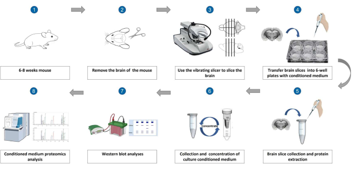
This article describes the basic procedures for (i) the isolation of brain slices, (ii) the collection of brain slices, (iii) the collection of brain slice cultures from conditional medium, (iv) western blot analyses, and (v) proteomic analysis (Figure 1).
Protocol
This protocol was approved and follows the animal care guidelines set by the Southern University of Science and Technology Animal Care and Use Committee (SUSTech-JY202112006). Adult male and female C57BL/6J mice (6-8 weeks old, 22-30 g) were used in this study. Mice were housed at 22-25 °C on a circadian cycle of 12 h of light and 12 h of darkness, with ad libitum access to food and water. The steps of the protocol are listed as follows.
1. Isolation of brain slices
- Prepare the surgical tools: ophthalmic bending tweezers, beakers (250 mL, 500 mL), filter paper, scissors, razor blade, single-sided blade, and brush pen. Place these tools in a foam box filled with crushed ice to cool.
- Open the valve for the mixed gas containing 95% O2 and 5% CO2. Clean the gas pipe head by placing it into a beaker of ultra-pure water.
- Prepare the ice-cold dissection buffer using the following components: 225 mM sucrose, 2.5 mM KCl, 1.25 mM NaH2PO4, 26 mM NaHCO3, 11 mM D-glucose, 5 mM L-ascorbic acid, 3 mM sodium pyruvate, 7 mM MgSO4, and 0.5 mM CaCl2. Adjust the pH to 7.3-7.4.
- Prepare the culture solution with the following components: 122 mM NaCl, 2.5 mM KCl, 1.25 mM NaH2PO4, 26 mM NaHCO3, 11 mM D-glucose, 7 mM MgSO4, and 0.5 mM CaCl2. Adjust the pH to 7.3-7.4, then filter and add 10% penicillin-streptomycin solution (P/S) into the culture solution.
- Oxygenate the culture solution and dissection solution for 30 min before sacrificing the mice. Break the razor blade to take half of the slice, and then mount it on the slicer. Install the base and tool holder of the slicer.
- Sacrifice the mice by cervical dislocation, or by using 2%-3% isofluorane to anesthetize.
- Use scissors to cut off the skull at the neck. Cut the skin in the middle of the skull to expose the whole skull.
- Use ophthalmic scissors to cut the skull along the median line and use curved eye forceps to cut the skull apart.
- Finally, use curved forceps to reach the base of the skull and cut off the base nerve; the entire brain can now be removed.
- Rapidly place the brain into an ice-cold dissection buffer. Use a sharp, sterilized, single-sided blade to cut away excess parts of the brain, including the bottom part.
- Stick the brain tissue into the tank using appropriate glue. Pour the slicing solution into the tank, ensuring that the brain tissue is covered, and continue to oxygenate.
- Adjust the parameters of the vibratome to slice the tissue into 300 µm thick sections. The speed and frequency of the slicer used in this study were 0.3 mm/s and 70 Hz, respectively.
- Use a brush to pick out the brain slices and place them into a dish with the culture solution. Then, transfer two to four slices onto each 30 mm, 0.4 µm pore size tissue culture insert in 6-well plates containing 1.5 mL of the culture solution, at a distance that does not allow the slices to grow into each other.
- Incubate the tissues at 37°C, 5% CO2, and 95% humidity for 14 h.
2. Brain slice collection
- Aspirate the culture medium from the coverslip and gently transfer the brain slice into the 1.5 mL tube with a brush.
- Add 200-400 µL of Hanks' balanced salt solution (HBSS) containing 1 mmol/L phenylmethylsulfonyl fluoride (PMSF) to the tube. Rigorously oscillate and mix to form a homogeneous solution using a tissue grinder. The frequency and time used in this study were 800 Hz and 60 s, respectively.
- Centrifuge the suspension at 700 x g for 10 min at 4 °C. Then, collect the supernatant into a new 1.5 mL centrifuge tube.
- Add 20-40 µL of 20% Triton X-100 to the supernatant and rotate the sample with a rocking shaker at 4°C for 1 h.
- Centrifuge the suspension at 13,000 x g for 20 min at 4 °C and collect the supernatant. Normalize all the brain slice samples to a protein concentration of 2 µg/µL using a bicinchoninic acid (BCA) protein assay kit.
- Mix the supernatant in 5x sodium dodecyl sulfate (SDS) loading buffer (10% SDS, 10 mM dithiothreitol [DTT], 250 mM Tris-HCl [pH 6.8], 30% [v/v] glycerol, and 0.05% [w/v] bromophenol blue dye) and boil the sample for 5 min at 95 °C.
- Freeze the prepared samples at -20 °C. They will subsequently be subjected to detection by electrophoresis and western blotting methods.
3. Collection of brain slice culture conditioned medium (CM)
- Aspirate the culture medium after 14 h and filter the CM using a 0.22 µm filter.
- Concentrate the conditioned medium up to 100 µL with a 10 kDa ultrafiltration centrifuge tube at 9,000 x g for 50 min at 4 °C. Normalize all medium samples to a protein concentration of 0.8 µg/µL using the BCA protein assay kit.
- Elute the conditioned medium in 5x SDS loading buffer and boil for 5 min at 95 °C.
- Freeze the prepared samples at -20 °C. They will subsequently be subjected to detection by electrophoresis and western blotting methods.
4. Western blot analyses
- Prepare sodium dodecyl-sulfate polyacrylamide gel electrophoresis (SDS-PAGE )gel as previously described10. Load the brain slice and medium sample onto precast SDS-PAGE gels. Separate the samples at a constant voltage of 80 V for 30 min initially, and then at a constant voltage of 120 V for 70 min.
- Transfer the proteins to polyvinylidene difluoride (PVDF) membranes and block with 5% bovine serum albumin (BSA) in tris-buffered saline-Tween 20 (TBST; 150 mM NaCl, 20 mM Tris, 0.1% Tween-20, pH 7.4) for 1 h.
- Add primary antibodies in the TBST as follows: mouse anti-sAPP-α (antibody dilution ratio of 1:5,000), rabbit anti-clusterin (antibody dilution ratio of 1:5,000), mouse anti-MAP2 (antibody dilution ratio of 1:5,000), mouse anti-GAPDH (antibody dilution ratio of 1:5,000), and mouse anti-actin (antibody dilution ratio of 1:5,000), and incubate overnight at 4 °C on a rocker.
- Wash the membrane three times with TBST for 15 min each.
- Add an appropriate secondary antibody in TBST buffer as follows: m-IgGκ BP-HRP (1:5,000 dilution), mouse anti-rabbit IgG-HRP (secondary antibody dilution ratio of 1:5,000). Incubate for 1-2 h.
- Wash the membrane three times using TBST for 15 min each.
- Scan and image the membranes using an appropriate imaging system.
5. Proteomic analysis
- In this study, collect the culture medium (CM) in accordance with step 3.1 and subsequently concentrate it to a final volume of 80 µL by using a 3 kDa concentration tube at 9,000 x g for 40 min at 4 °C. Store the CM sample on ice, awaiting mass spectrometry analysis.
- Conduct the proteomic detection and analysis methods for samples following the protocols as described in previous papers11,12,13.
Results
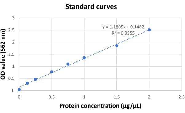
To quantify the secretion of extracellular proteins in brain slices, we examined protein concentrations of the brain slice samples and conditioned medium samples through BCA assay experiments. Brain slice samples and conditioned medium samples all had high protein concentrations (Table 1 and Figure 2). We found that a large number of extracellular proteins were secreted into the medium, and the average protein concentrations in the conditioned medium were calculated to be 1.78 µg/µL. The brain slices remained alive after 14 h of incubation, and the average protein concentration of the brain slices was 4.5 µg/µL. It should be noted that once the brain slices died, the protein concentrations detected in the medium and slices were extremely low.
To validate that a large quantity of important neurosecretory proteins is produced in the CM using this method, we performed a proteomic analysis on the collected CM and detected a total of 2,390 proteins. To further analyze the abundance of key neuronal proteins in the collected secretome, we conducted a Venn diagram analysis of the data in comparison with a previously published dataset of the brain secretome and a dataset of mouse cerebrospinal fluid (CSF) proteomes14,15,16. From the results, we found that the number of proteins obtained from mouse brain CSF in the previous study was only 616, which is nearly four times lower than the number of proteins obtained from brain slice CM. At the same time, we detected 1,300 neurosecretory proteins in the brain slice CM, while only 140 neurosecretory proteins were detected in the CSF. This indicates that our brain slice CM obtained not only neurosecretory proteins, but also non-neurosecretory proteins. It is worth noting that the number of neurosecretory proteins extracted from the brain slice CM is four times higher than that from the CSF extraction method (Figure 3). In addition, a large number of important neurosecretory proteins can be found in brain slice CM that are absent in CSF, such as neuroligin (NLGN) family proteins. Detailed information about these neurosecretory proteins can be found in Table 2, which demonstrates that this method collects a rich amount of the critical brain secretome and has a wider range of applications, making it suitable for the analysis of multiple types of tissues.
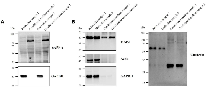
To further examine the formation of several key neuronal and synaptic secreted proteins in the medium, such as microtubule-associated protein (MAP2), amyloid precursor protein (APP), neuroligin, and neurexin, we performed western blot experiments on the brain slice and CM samples. The antibodies used in this experiment could only recognize the extracellular APP protein (sAPP); intracellular APP could not be detected. MAP2 and clusterin antibodies could recognize both intracellular and extracellular proteins simultaneously. The western blot results show that sAPP protein is only found in the CM (Figure 4A). MAP2 and clusterin can be detected both intracellularly and extracellularly, indicating that the APP and MAP2 protein can also be isolated in the CM (Figure 4B). The results of protein composition analysis of the culture medium also showed the expression of MAP2, clusterin, and APP proteins (Table 2). Besides several critical neural protein markers in the culture medium, we used two non-neural proteins as controls: GAPDH and actin. We found that GAPDH and actin were barely detectable in the CM samples, but were highly expressed in the brain slice samples. This indicates that the brain slice CM completely separated secreted and non-secreted proteins; secreted proteins were only significantly present in the medium, reducing the interference from non-secreted proteins in the brain slices. Two sets of replicate experiments were performed simultaneously, and both replicates showed a high degree of similarity (Figure 4). These data indicate that many key neuronal and synaptic proteins could be found in the CM, and that isolation of the brain secretome using ex vivo brain slice cultures is feasible and reliable.

Figure 1: Schematic diagram for isolation of the brain secretome from ex vivo brain slice cultures. (1) Choosing a 6-8-week-old mouse. (2) Removal of the brain of the mouse. (3) Use of the vibrating slicer to slice the brain. (4) Transferring the brain slices into 6-well plates with conditioned medium. (5) Brain slice collection and protein extraction. (6) Collection and concentration of culture conditioned medium. (7) Western blot analyses. (8) Conditioned medium proteomics analysis. Please click here to view a larger version of this figure.

Figure 2: BCA standard curves. Concentration and absorbance linear relationship of the BSA standard. Protein concentration range of 0-2.5 µg/µL and an optical density (OD) value range of 0-3. All the analyses are run in duplicate. Please click here to view a larger version of this figure.

Figure 3: Proteomic analysis of the conditioned medium samples. Venn diagram of proteins from brain slice CM (n = 2390), proteins from the brain secretome (n = 1,252) and proteins from mouse CSF (n =616), and a histogram of the numbers of these three types of proteins. Please click here to view a larger version of this figure.

Figure 4: Western blot analysis of the brain slice samples and conditioned medium samples. Total brain slice lysates and concentrated CM were isolated and western blotting was applied. (A) Western blot analyses of the brain slice samples and conditioned medium samples by using sAPP-αantibody (1:1,000 diluted) and GAPDH antibody (1:1,000 diluted), respectively. (B) Western blot analyses of the brain slice samples and conditioned medium samples using MAP2 antibody (1:1,000 diluted), clusterin antibody (1:1,000 diluted), GAPDH antibody, and actin antibody (1:1,000 diluted) respectively. The first two lanes are replicates of brain slice samples, and the last two lanes are replicates of conditioned medium samples. Please click here to view a larger version of this figure.
Table 1: BCA analysis of brain slice samples and conditioned medium samples. Total brain slice lysates and concentrated CM were isolated and prepared for BCA. Quantitative analysis of protein concentration in three sets of repeated brain slice and conditioned medium samples, according to the OD value of the sample detected at 562 nm. Please click here to download this Table.
Table 2: The expression levels of the critical brain secretome in conditioned medium samples. Proteomic analysis of the brain slice culture medium. Relevant information on 22 key proteins in the brain secretome, identified by proteomic data. Please click here to download this Table.
Discussion
The brain secretome refers to the collection of signaling molecules, known as neurosecretory or neuropeptide products, that are released by neurons or glial cells into the extracellular environment. The brain secretome holds critical functions in many biological and physiological processes in the nervous system17,18. Understanding the brain secretome and its functions is important for gaining insight into the mechanisms underlying brain function and disorders. Moreover, brain secretory proteins demonstrate potential value as novel therapeutic targets for advancing drug development or as biomarkers to aid in disease diagnosis and treatment19,20. The extraction of the brain secretome has always posed a challenge due to its cumbersome and time-consuming procedure and high failure rate in experiments. Building on the work of Pandamooz et al. with cultured spinal cord sections of adult rats in vitro, the focus of this experiment was primarily on establishing a spinal cord slice culture model and extracting proteins related to spinal cord tissue, rather than exploring the extraction and separation of neuronal secretory proteins21. In this protocol, we solved the above problems and isolated the brain secretome by using ex vivo brain slice cultures. There are some practical advantages of this method: firstly, it provides an excellent model for the study of the brain secretome; secondly, it takes less time and has lower experimental costs compared to the traditional isolation method; lastly, the method of extracting the secretome is not difficult to operate and would be feasible for even the most inexperienced researchers.
In this experiment, it is critical to maintain the activity of brain slices in order to ensure accurate results during protein collection. Practicing dissection and sectioning operations is important for the quality and survival of brain slices. During the process of using the vibrating slicer, it is important to ensure that the dissection solution is a mixture of ice water, which will improve the quality and activity of brain slices. Moreover, removing the mouse brain as quickly as possible is also a critical step in maintaining the activity of brain slices. Finally, it is necessary to ensure the integrity of the brain slices is preserved and that the slices are not crushed when they are being transferred to the 6-well plates.
During the brain slice culture process, various drugs can be added to the medium depending on the experimental needs; however, the exact drug concentrations remain to be explored. In addition, excessively long-term cultures can lead to the death of brain slices. In this experiment, we found that the brain secretome can be detected in the medium after 14 h of culture. The choice of time depends on the expression level of the protein to be collected. When collecting proteins with lower expression levels, it is appropriate to extend the cultivation time of brain slices. Based on our preliminary experiments, neurons secrete a low amount of protein before 12 h of cultivation, but the survival rate of brain slices is low after 18-24 h of cultivation, and the concentration of whole protein in dead brain slices is very low. Therefore, collecting culture medium from 12-18 h would provide the best results in terms of both the protein concentration of neuron-secreted protein and the quality of brain slices.
There were mainly two different methods available for the extraction of the brain secretome: cell culture-based methods22 and collection from cerebrospinal fluid1. Isolation of the brain secretome from brain slices holds some advantages compared to cell culture-based methods. Firstly, brain slices provide a more in vivo-like environment, as they maintain the native architecture and connectivity of the tissue, which can result in a more accurate representation of the brain secretome. Secondly, isolation of the brain secretome from brain slices has a shorter experimental time period and higher availability. However, there are some variabilities between brain slices, which can influence the results of the extraction. In comparison to collection of the brain secretome from cerebrospinal fluid, it is easier and more convenient to extract the secretome from brain slices, although it is also important to note that brain slices may only be available from specific regions of the brain and the number of slices obtained may be limited, which can impact the robustness of the data obtained.
One of the limitations of this protocol is that the medium is enriched using a 10 kDa ultrafiltration centrifuge tube so that proteins with molecular weights below 10 kDa are discarded. Comparatively, this method greatly increased the concentration of residual protein. Another weakness of this method is that the obtained proteins not only include neurosecretory proteins but also non-neurosecretory proteins; however, this also means that the method can be used to study other neuroendocrine proteins in the brain. In addition, this method can also be considered for extracting secreted proteins from other types of tissues. Moreover, the expression of each protein is different after a period of culture, so it is necessary to adjust the experimental conditions according to the needs of the experiment.
Taken altogether, we have presented a detailed protocol for isolating the brain secretome. We have also proved that the brain slices and the conditioned medium still contain a large number of critical brain secretome proteins after a few hours of culture. The method is rapid and simple, allowing it to be widely applied in the fields of biology, pharmacology, and medicine.
Disclosures
The authors state that they do not have any conflicts of interest.
Acknowledgements
This research was supported by Shenzhen Clinical Research Center for Mental Disorders (20210617155253001), Shenzhen Fund for Guangdong Provincial High-level Clinical Key Specialties (SZGSPO13), Shenzhen Science and Technology Innovation Committee (JCYJ20200109150700942), Key-Area Research and Development Program of Guang Dong Province (2019B030335001), and Shenzhen Key Medical Discipline Construction Fund (SZXK042) We are grateful for the assistance of Guangdong Provincial Key Laboratory of Advanced Biomaterials.
Materials
| Name | Company | Catalog Number | Comments |
| 0.22 μm Non-Sterile Millex Syringe Filters with PES Membrane | Millipore | SLGPR33RB | |
| 1,4-Dithio-DL-threitol (DTT) | Merck (Sigma Aldrich) | DTT-RO | |
| 6-well plates | CORNING | 3516 | |
| Actin antibody | Cell Signaling Technology | #3700 | |
| APP Antibody | Cell Signaling Technology | 2452 | |
| BioRad Mini-Protean TGX precast gels | BioRad | #1610185 | |
| Bovine serum albumin (BSA) | Merck (Sigma Aldrich) | 10735094001 | |
| Bromophenol Blue | Merck (Sigma Aldrich) | B0126 | |
| Brush pen | Deli | 1.00008E+11 | |
| CaCl2 | Merck (Sigma Aldrich) | C1016 | |
| CF3COOH | Merck (Sigma Aldrich) | 302031 | |
| CH3CN | Merck (Sigma Aldrich) | 34851 | |
| Clusterin antibody | Cell Signaling Technology | #42143 | |
| D-Glucose | Merck (Sigma Aldrich) | G8270 | |
| Easy-nLC1200 | Thermo Fisher Scientific | Easy-nLC1200 | |
| Filter paper | ROHU | ROHU-DLLZ00105 | |
| GAPDH antibody | Cell Signaling Technology | # 5174S | |
| Glycerol | Merck (Sigma Aldrich) | G5516-100ML | |
| Hanks' Balanced Salt Solution (HBSS) | Invitrogen (Gibco) | 14175095 | |
| Human/Rat NLGN2 Antibody | R&D Systems | AF5645 | |
| ICH2CONH2 | Merck (Sigma Aldrich) | I6125 | |
| Immun-Blot PVDF membranes | BIO-RAD | #1620177 | |
| KCl | Merck (Sigma Aldrich) | P3911 | |
| L-Ascorbic Acid | Merck (Sigma Aldrich) | A92902 | |
| Map2 antibody | Cell Signaling Technology | # 4542S | |
| MgSO4 | Merck (Sigma Aldrich) | M7506 | |
| m-IgGκ BP-HRP | SANTA CRUZ BIOTECHNOLOGY | sc-516102 | |
| Millicell-CM 0.4 μm | Millipore | PIHP03050 | |
| Millipore AmiconUltra-0.5ML | Millipore | UFC5010 | |
| mouse anti-rabbit IgG-HRP | SANTA CRUZ BIOTECHNOLOGY | sc-2357 | |
| NaH2PO4 | Merck (Sigma Aldrich) | S0751 | |
| NaHCO3 | Merck (Sigma Aldrich) | S5761 | |
| NH4HCO3 | Merck (Sigma Aldrich) | A6141 | |
| Parenzyme | Thermo Fisher Scientific | R001100 | |
| Penicillin-Streptomycin Solution(P/S) | Invitrogen (Gibco) | 15070063 | |
| Pierce BCA Protein Assay Kit | Thermo Fisher Scientific | 23225 | |
| QEactive | Thermo Fisher Scientific | IQLAAEGAAPFALGMAZR | |
| Rabbit Anti-Goat IgG (H&L)-HRP Conjugated | Easybio | BE0103-100 | |
| Razor blade | BFYING EAGLE | HX-L146-1H | |
| Sodium chloride NaCl | Merck (Sigma Aldrich) | S6150 | |
| Sodium dodecylsulfate (SDS) | Merck (Sigma Aldrich) | V900859 | |
| Sodium Pyruvate | Merck (Sigma Aldrich) | P2256 | |
| SpeedVac SRF110 Refrigerated Vacuum Concentrator (German) | Thermo Fisher Scientific | SRF110P2-115 | |
| Sucrose | Merck (Sigma Aldrich) | G8270 | |
| Tissue grinder | SCIENTZ | SCIENTZ-48 | |
| Tris (Hydroxymethyl) | Merck (Sigma Aldrich) | TRIS-RO | |
| TritonX-100 | Merck (Sigma Aldrich) | T8787 | |
| Tween-20 | Merck (Sigma Aldrich) | P1379 | |
| Vibratory slicer | Leica | Leica VT 1000S |
References
- Martín-de-Saavedra, M. D., et al. Shed CNTNAP2 ectodomain is detectable in CSF and regulates Ca2+ homeostasis and network synchrony via PMCA2/ATP2B2. Neuron. 110 (4), 627-643 (2022).
- Jha, M. K., et al. The secretome signature of reactive glial cells and its pathological implications. Biochimica et Biophysica Acta. 1834 (11), 2418-2428 (2013).
- Maćkowiak, M., Mordalska, P., Wędzony, K. Neuroligins, synapse balance and neuropsychiatric disorders. Pharmacological Reports. 66 (5), 830-835 (2014).
- Suzuki, K., et al. Activity-dependent proteolytic cleavage of neuroligin-1. Neuron. 76 (2), 410-422 (2012).
- Venkatesh, H. S., et al. Targeting neuronal activity-regulated neuroligin-3 dependency in high-grade glioma. Nature. 549 (7673), 533-537 (2017).
- Shen, F., et al. Proteomic analysis of cerebrospinal fluid: toward the identification of biomarkers for gliomas. Neurosurgical Review. 37 (3), 367-380 (2014).
- Suk, K. Combined analysis of the glia secretome and the CSF proteome: neuroinflammation and novel biomarkers. Expert Review of Proteomics. 7 (2), 263-274 (2010).
- Pegg, C. C., He, C., Stroink, A. R., Kattner, K. A., Wang, C. X. Technique for collection of cerebrospinal fluid from the cisterna magna in rat. Journal of Neuroscience Methods. 187 (1), 8-12 (2010).
- Bossu, J. L., Capogna, M., Debanne, D., McKinney, R. A., Gähwiler, B. H. Somatic voltage-gated potassium currents of rat hippocampal pyramidal cells in organotypic slice cultures. The Journal of Physiology. 495, 367-381 (1996).
- Hirano, S. Western blot analysis. Methods in Molecular Biology. 926, 87-97 (2012).
- Davey, J. R., et al. Integrated expression analysis of muscle hypertrophy identifies Asb2 as a negative regulator of muscle mass. Journal of Clinical Investigation Insight. 1 (5), e85477 (2016).
- Ren, H., et al. Identification of TPD52 and DNAJB1 as two novel bile biomarkers for cholangiocarcinoma by iTRAQ-based quantitative proteomics analysis. Oncology Reports. 42 (6), 2622-2634 (2019).
- Li, J., et al. Identification and characterization of 293T cell-derived exosomes by profiling the protein, mRNA and MicroRNA components. PLoS One. 11 (9), 0163043 (2016).
- Kuhn, P. H., et al. Systematic substrate identification indicates a central role for the metalloprotease ADAM10 in axon targeting and synapse function. eLife. 5, e12748 (2016).
- Chiasserini, D., et al. Proteomic analysis of cerebrospinal fluid extracellular vesicles: a comprehensive dataset. Journal of Proteomics. 106, 191-204 (2014).
- Dislich, B., et al. Label-free quantitative proteomics of mouse cerebrospinal fluid detects β-site APP cleaving enzyme (BACE1) protease substrates in vivo. Molecular and Cellular Proteomics. 14 (10), 2550-2563 (2015).
- Chenau, J., Michelland, S., Seve, M. Secretome: definitions and biomedical interest. La Revue de Médecine Interne. 29 (7), 606-608 (2008).
- Assunção-Silva, R. C., et al. Exploiting the impact of the secretome of MSCs isolated from different tissue sources on neuronal differentiation and axonal growth. Biochimie. 155, 83-91 (2018).
- Ardianto, C., et al. Secretome as neuropathology-targeted intervention of Parkinson's disease. Regenerative Therapy. 21, 288-293 (2022).
- Willis, C. M., Nicaise, A. M., Peruzzotti-Jametti, L., Pluchino, S. The neural stem cell secretome and its role in brain repair. Brain Research. 1729, 146615 (2020).
- Pandamooz, S., et al. Modeling traumatic injury in organotypic spinal cord slice culture obtained from adult rat. Tissue and Cell. 56, 90-97 (2019).
- Peixoto, R. T., et al. Transsynaptic signaling by activity-dependent cleavage of neuroligin-1. Neuron. 76 (2), 396-409 (2012).
Reprints and Permissions
Request permission to reuse the text or figures of this JoVE article
Request PermissionThis article has been published
Video Coming Soon
Copyright © 2025 MyJoVE Corporation. All rights reserved