Method Article
An Integrated (Microscopic/Endoscopic) Dissection Ear Surgery Course
In This Article
Abstract
Traditionally, otologic surgical training consisted of microscopic cadaveric dissections. However, during the last decades the endoscope has significantly changed the surgical perspective in the otologic field. Thus, the modern ear and lateral skull base surgeon should master the entire spectrum of endoscopic and microscopic approaches, with the aim of tailoring the procedure and guaranteeing the best possible functional outcome. This work proposes a step-by-step guided and illustrated dissection course, including indications for the setup of the cadaver lab and the integration of the microscope and endoscope to enhance the use of both instruments. The alternation of the endoscope and microscope allows the novice to train the correct handling of the instruments in the surgical field under both optical views. This aspect is of utmost importance since it is not advisable to start off a technique without practicing the other one, as both are important and complementary in the modern otologic surgery setting.
Introduction
Traditionally, otologic surgical training consisted of microscopic cadaveric dissection in order to develop both transcanal and transmastoid procedures. However, during the last decades the endoscope has significantly changed the surgical perspective. Nowadays, a consistent number of patients can benefit from minimally invasive endoscopic ear surgeries1,2,3,4,5,6. Thus, the modern otologic surgeon should master the entire spectrum of both endoscopic and microscopic approaches, with the aim of tailoring the procedure to the disease and the patient and guaranteeing the best possible functional outcome.
Dissection courses are notably expensive due to the high prices of the cadaveric specimens and the need of up-to-date technological equipment (e.g., microscope, endoscope, high-definition cameras and monitors, high-speed drills, piezoelectric devices, etc.). Moreover, the availability of fresh human cadavers is limited and might be further restricted by financial and regulatory issues. Therefore, it would be wise to maximize the efficiency of the resources to perform any possible microscopic and endoscopic dissection step on one single specimen.
Here, we present a protocol that systematizes all the steps for a comprehensive middle ear and lateral skull base dissection course that enhances the trainee's experience on different surgical procedures, with both the microscope and the endoscope. This work proposes a step-by-step guided and illustrated dissection course, including indications for the setup of the cadaver lab. This innovative approach consists in the integration of the microscope and the endoscope, which are repeatedly alternated throughout the dissection course. By doing this, the trainee can perform a stepwise dissection that preserves the anatomical landmarks needed for the further surgical steps, exploiting the use of both instruments. This protocol derives from the wide experience of our team in teaching both surgical approaches in gross anatomy lab. In fact, this method has been applied for years during both national and international dissection courses.
Protocol
The following protocol follows the guidelines of our institution's human research ethics committee. The ethics committee approved the protocol.
1. Preparation of the specimen
- Position a vacuum mattress on the dissection table and cover it with a sterile and resorbable blanket. Lay the anatomical specimen in a surgical position on the mattress with the head rotated to the contralateral side.
- Cover the specimen with a blanket sparing the external ear and retroauricular area, just as in the operating theater.
- Consider the possibility to preliminarily scan the specimen through cone-beam or traditional CT, for radiologic guidance during dissection.
NOTE: Please see the Table of Materials for a complete list of the equipment required.
2. Getting started
- Adopt a comfortable sitting position in front of the specimen, adjust the endoscope screen and the microscope.
- Perform a white balance of the camera.
- Perform the following under microscopic and/or endoscopic vision (Table 1).
NOTE: The letter at the beginning of each paragraph, indicating the preferred tool to perform dissection (microscope = M; endoscope = E). M/E indicates that the choice of the technique to adopt is up to the trainee in that step.
3. M: Retroauricular skin incision
- Use a scalpel to perform standard retroauricular skin incision about 1 cm behind the retroauricular sulcus, covering 180° degrees of the external auditory canal (EAC).
NOTE: The autologous fascia of the temporalis muscle can be harvested for further steps (e.g., myringoplasty, scutum reconstruction). - Denude the bony area behind and above the auricle by means of a periosteal elevator to place the self-retaining retractors before the following steps. It is of particular importance to skeletonize the zygomatic root above the EAC in order to perform the epitympanotomy in a later step.
4. M: Cortical mastoidectomy (Figure 1)
- General recommendations about drilling
- Start the dissection using the largest burr possible, since small burrs have higher penetration potential into the bone and can be dangerous if not adequately controlled.
- Perform most of the bone work with cutting burrs. Reserve diamond burrs for working near delicate structures (i.e., facial nerve, sigmoid sinus, middle cranial fossa dura).
- Hold the drill like a pen and try to apply the drill in a tangential rather than perpendicular direction to the structures being drilling to use the burr's equator instead of its tip.
- Apply minimal or no pressure during drilling.
- Start drilling from most dangerous structures, when identified, to the least dangerous ones.
- To help stabilize the surgeon's hand and drill, place the small finger on the head of the specimen. Another useful trick is to place the sucker between a structure of importance and the burr. In this way, if the control of the burr is lost, it may hit the sucker instead of the structure of interest.
- Begin drilling of the mastoid cortex using a large cutting burr identifying the middle cranial fossa (MCF) dura, approximately at the level of the temporalis ridge. The signal that the level of the dura is close is usually provided by the appearance of the different dural color through the bone and by the change in the burr's noise to a high-pitched sound.
- Start drilling at the expected level of the sigmoid sinus, which is on an oblique line connecting the posterior edge of the temporal ridge to the tip of the mastoid bone.
- Join the two lines of drilling created by drilling the posterior tangent of the posterior EAC wall, thus creating the so-called "triangle of attack".
- Drill out the bone in the center of the triangle by deepening the cavity evenly and gradually. Saucerize the cavity to provide the optimal visualization. Thin out the bone over the sigmoid sinus and the MCF dura with a diamond burr (3-5 mm diameter).
- Follow the MCF plane to identify and open the antrum, achieving an adequate view on the lateral semicircular canal (LSC).
- Tilt the specimen away from the surgeon to identify the body and the short process of the incus. Take care to avoid touching it with rotating burr to preserve the ossicular chain intact for further steps.
5. M/E: Myringotomy (optional)
- Advance a myringotomy knife towards the eardrum and perform a radial incision at the antero-inferior tympanic quadrant. The incision must be large enough to accommodate a ventilation tube.
- Pick a standard Donaldson type tube with Hartmann forceps, introduce in the EAC and place on the eardrum close to the myringotomy incision.
- Using a 1.5 mm, 45° hook, rotate the inner flange through the myringotomy incision so that the tube straddles the tympanic membrane.
6. M/E: Tympanomeatal flap and middle ear anatomy exploration (Figure 2 and 3)
- Raise the tympanomeatal flap by means of microscope or endoscope, as preferred by the surgeon. A 0° endoscope is adequate for this step. Adjust the endoscope screen to achieve a comfortable neck position while dissecting.
- Clean the EAC from ear wax and any desquamated skin debris with the suction tube and Hartman forceps, as needed.
- Trim EAC hair with a pair of small scissors to prevent the endoscope to become dirty during every passage through the EAC. Keep the scissors' blades perpendicular to the EAC surface, to effectively cut the hairs and not to damage the skin.
- Incise the EAC skin with a round knife from the 11 o'clock to the 6 o'clock positions.
- Gently dissect the skin of the EAC with an elevator, from lateral to medial, until the fibrous annulus and the Prussak space is reached.
- Dissect the pars flaccida of the lateral process of the malleus by pulling it inferiorly with Hartmann forceps. Elevate the tympanomeatal flap, keeping it attached to the handle of the malleus.
- To complete the flap elevation, free the handle of the malleus from its adhesion to the eardrum, by pulling it using a Hartmann forceps or by dissecting it with a hook from superiorly to inferiorly.
- At the end, detach the eardrum from the umbus by cutting it with microscissors.
- Explore the middle ear anatomy by means of 0° and 45° endoscope, as already described in a previous protocol7.
- In the epitympanum, identify the malleus (neck, short process, manubrium and umbo; the incus (body, long process, lenticular process, incudostapedial joint); the epitympanic diaphragm (anterior and lateral malleolar ligaments, lateral incudomalleolar ligamental fold); the anterior and posterior spine; the chorda tympani8; the tympanic isthmus and ventilation patterns to the antrum; the cochleariform process; the tensor tympani muscle; the tendon and bony canal; and the tensor fold.
- In the mesotympanum, identify: the stapes (head, anterior and posterior crus, footplate); the tendon of stapedial muscle; the pyramidal eminence; the promontory bone; and the Jacobson nerve with inferior tympanic artery.
- In the retrotympanum, identify: the facial sinus9; the posterior sinus; the ponticulus; the sinus tympani10; the subiculum; the styloid eminence; the subtympanic sinus11; the fustis bone; the round window niche with tegmen, anterior and posterior pillar; the round window membrane; the subcochlear canaliculus12; and the finiculus.
- In the hypotympanum, identify hypotympanic cells and estimate localization of the jugular bulb13,14 if not visible.
- In the protympanum, identify: the protiniculus15; the internal carotid artery (ICA); and the eustachian tube (ET).
7. M/E: Myringoplasty (optional)
- Refresh the perforation's edges (e.g., myringotomy hole) with a knife slightly enlarging the perforation. Measure the perforation with a bended hook. Pull the tympanomeatal flap anteriorly onto the anterior aspect of the EAC.
- Remodel the chosen graft (e.g., temporalis muscle fascia, heterologous membrane) according with the perforation's size. Then, place it in the EAC and position it over the handle of the malleus and under the anterior lip of the perforation, in contact with the eardrum.
- Pack the tympanic cavity, especially the anteroinferior area, with gelfoam soaked with water, to sustain the graft.
NOTE: The tympanomeatal flap and the graft can be repositioned to complete the step.
8. M: Epitympanotomy
- Start from the posterior position, and choose a burr of sufficient size to fit into the space between the MCF dura and the superior wall of the EAC. Drill from the medial to the lateral direction, taking care not to touch the underlying ossicular chain.
NOTE: The anterior extent of the epitympanotomy should be sufficient to expose the anterior epitympanic space, lying anteriorly to the cog.
9. M: Posterior tympanotomy
- Use a small diamond burr (1-2 mm diameter) to open the facial recess, starting from bone below the short process of the incus (below the buttress) up to the chorda tympani. When performing this step, take care to not drill too far anterolaterally, jeopardizing the anulus and the eardrum.
- Finish the posterior tympanotomy once the triangle between the buttress. Drill out the chorda tympani and the mastoid portion of the facial nerve (FN), achieving an adequate access to the retrotympanum and the round window niche (e.g., cochlear implant).
- Tailor the inferior extension of the posterior tympanotomy and extend over the chorda tympani to improve the visualization of the middle ear cleft. To widen the posterior tympanotomy (extended facial recess), transect the chorda along the FN and following the annulus, all the bone anterior to the FN, inferior to the annulus plane and lateral to the jugular bulb and ICA is removed.
10. M: Decompression of the mastoid portion of the facial nerve (optional)
- Identify the digastric ridge by drilling the pneumatization of the mastoid tip. The FN is found in an anteromedial position with respect of it.
- Start identification of the mastoid segment of FN with a large cutting burr, which is moved parallel to the FN, whose position can be estimated by the position of the LSC and the short process of the incus.
- Once the whole segment of the FN can be seen through the bone, use a diamond burr (3-4 mm diameter) to skeletonize to a total of 270° of the FN canal.
- Remove the last shell of thin bone covering the FN using a curved hook, thus uncovering the FN from the second genu up to the stylomastoid foramen.
- Using a new Beaver knife with the sharp border facing away from the nerve, incise the perineural sheath of the nerve, completing the decompression of the mastoid portion of the FN.
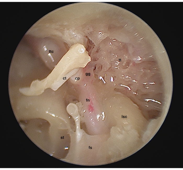
11. E: Atticotomy and ossicular chain removal (Figure 4)
- Remove the lateral wall of the epitympanum (scutum) with the curette, the drill or piezosurgery, if available. Beyond the instrument, be careful not to disarticulate the incudo-malleolar joint. Start atticotomy from the free inferior and posterior bony edge of the EAC, and progressively extend upward, to completely expose the incudo-malleolar joint.
- Disarticulate the incudo-stapedial joint with round knife, and gently remove the incus. Identify the LSC, the tympanic tract of the FN, the geniculate ganglion area, the cochleariform process, the tegmen tympani (already thinned and smoothened through the microscopic epitympanotomy).
- Use the malleus punch to cut the malleus neck and displace the malleus head. Then, use Bellucci's scissors to cut the tendon of the tensor tympani muscle and remove the malleus handle.
12. M/E: Ossiculoplasty (optional)
- Use either the incus or the malleus head. Under microscopic view, use the drill to model the graft (remove the long and short process of the incus or smoothen the edges of the malleus head). In both cases, perform a small hole (around 1 mm - same as the burr diameter), to fit the stapes head.
- Under endoscopic view, place the carved graft in the ear canal, and with the hook, gently move it to position it on the stapes, making sure that good contact is maintained.
NOTE: Moreover, it is possible to perform ossicular chain reconstruction by means of titanium prosthesis, if available, to try a device often employed in real-life situation.
13. E: Tympanic facial nerve decompression and access to the geniculate ganglion2
- Remove the cochleariform process with the curette, free the tensor tympani muscle from its canal and displace it towards the ET (you can also cut it).
- Using the curette or the round knife, gently peel off the Fallopian canal covering the whole tympanic tract of the FN (from the cochleariform process to the area of the second genu). This bone is very thin and sometimes the nerve is already dehiscent.
- Drill the tegmen tympani to expose the dura mater of the MFC. Check the microscopic view of the tegmen tympani from the retroauricolar approach: imagine the line of dural exposure in the posterior cranial fossa (triangle of attack) continuing anteriorly with the dura of the tegmen antri and tegmen tympani.
- Remove any bone between the most proximal part of the tympanic FN and the anterior dura, to expose the geniculate ganglion and the greater superior petrosal nerve (GSPN).
- Gently stretching the completely freed FN with the aid of a bended dissector, identify the first genu and the proximal part of the labyrinthine segment of the FN, heading towards the internal auditory canal, in an inferior and medial direction (respect to the plane of the tympanic segment).
- Note that parallel and anterior to the geniculate ganglion is the tympanic nerve of Jacobson, which runs towards the MCF dura with an inferior-to-superior path.
14. M: Endolymphatic sac decompression (Figure 5)
- Complete the skeletonization of the sigmoid sinus; the drilling on its medial wall should be deep enough into the bone of the posterior cranial fossa (PCF).
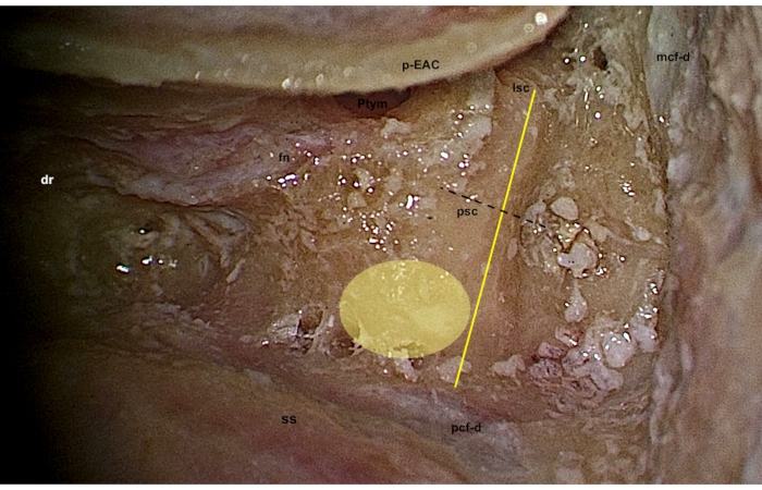
- Complete the skeletonization of the posterior semicircular canal (PSC), trying not to open it, and identify the Donaldson's line: this line passes through the LSC, bisecting the PSC, pointing at the sigmoid sinus .
- Localize the endolymphatic sac as a thickening of the dura of the PCF inferior and medial to the Donaldson's line, so that the area to be drilled is between the PSC and the sigmoid sinus, and between the superior petrosal sinus and the jugular bulb, towards the retrofacial recess.
- The location of the sac could be variable: look for a white pearl thickening of the dura or a fine hypervascular net on the dura. Use the diamond burr to slightly peel off the bone covering on the sac.
- Identify the endolymphatic duct as an apical extension towards the PSC in the postero-superior part of the sac, gently pushing on the sac itself with a dissector.
- Open the sac with a sickle or round knife and identify its medial wall.
15. M: Retrofacial approach
- To simulate retrofacial approach, drill the retrofacial cells, staying parallel to the posterior wall of the third tract of the FN. The limits of this approach are the PSC and the cochlea superiorly, the dura of the PCF posteriorly (with the endolymphatic sac already opened) and the jugular bulb antero-inferiorly.
16. E: Round window niche anatomy and transpromontorial approach to the internal acoustic canal (Figure 6)
- To achieve adequate surgical area of maneuvering, use the drill or piezosurgery to skeletonize: the temporo-mandibular joint (anterior superficial limit), the ICA in the protympanic region (anterior deep limit) and the jugular bulb in the hypotympanic region (inferior deep limit). The other landmarks (MCF dura -superior superficial limit, the second segment of the FN - superior deep limit, and the third segment of the FN - the posterior limit) have already been skeletonized16,17,18.
- Using the curette, remove the tegmen of the round window to identify the round window membrane, and to study the relationship with the subcochlear canaliculus and the fustis.
- After cutting the stapedial tendon, remove the stapes. It is possible to identify the spherical recess by means of 0° endoscope.
- Widen the oval window opening inferiorly towards the round window using the curette and the area of the vestibule is enlarged.
- Identify three landmarks on the medial wall of the vestibule (vestibular bony labyrinth): the lowest is the spherical recess (termination of the inferior vestibular nerve fibers), the highest is the elliptical recess (termination of the superior vestibular nerve fibers) and in between the two recesses, the vestibular crest could be identified. Note that the bone of the promontorial region is thick, thus some strength should be applied to remove it.
- Remove the round window membrane with a hook, and identify the scala vestibuli, scala tympani, and the spiral lamina in between.
- Progressively drill along the promontorium with a small diamond burr, on the lateral surface of the cochlea, to expose basal, middle and apical cochlear turns and the modiolus. To avoid damage, follow the dissection lines as parallel to the turns of the cochlea, gently drilling the bony surface.
- To get access to the medial wall of the cochlear bony labyrinth, remove the bony septa in between the cochlear turns and the modiolus.
- Drill to reach the fundus of the internal auditory canal and to possibly see the entrance of the cochlear nerve into it (sometimes it is not possible to clearly distinguish the cochlear nerve after extensive drilling on the modiolus). Following the labyrinthine segment of the FN, identify the intrameatal portion of the FN and observe the relationship with the cochlear nerve posterior to it (the two nerves draw a "Y" figure in this area)
17. M: Canal wall down (CWD) tympanoplasty
- To perform CWD tympanoplasty, most of the steps have already been performed (cortical mastoidectomy, epitympanotomy, thinning of the posterior bony EAC, skeletonization of the FN). Drill the posterior wall of the EAC with a cutting burr. Flatten the anterior buttress (bony crest between the posterior wall of the EAC and the tegmen tympani), as well as the posterior (incus) buttress, if kept so far.
18. M: Labyrinthectomy and translabyrinthine approach to the internal auditory canal (IAC) (Figure 7)
- Complete the skeletonization of the sino-dural angle. Remove the shell of bone over the sigmoid sinus and the dura posterior to it using a large diamond burr. The MCF dura is similarly uncovered.
NOTE: Understanding the relationship between the SCCs is fundamental for labyrinthectomy. The labyrinth dissection starts with the skeletonization of the LSC, along its major axis, to reach the so called "blue line", which is the appearance of the membranous labyrinth inside it. The procedure similarly continues with the drilling of the PSC and the superior semicircular canal (SSC). The SSC runs posterior and medial to create the common crus, connecting with the PSC. The latter must be drilled until the most infero-anterior portion, close to the third tract of FN. - Identify of the subarcuate artery in the hard bone at the center of the circle drawn by the SSC.
- Open the vestibule, which is situated in front of the common crus, and widen this region with progressive drilling to remove all the membranous labyrinth (vestibule and SCCs).
- Drill the bony cover of the most medial part of the dura of the PCF and MCF that are now reachable by the drill.
- Identify the margins of the IAC: the superior margin is a line between the subarcuate artery and the SSC ampulla, running between the FN anteriorly and the sino-dural angle posteriorly; the inferior margin, parallel to the superior one, is marked by the line between the PSC ampulla antero-superiorly (area of the retrofacial cells) and the jugular bulb antero-inferiorly, towards the PCF dura posteriorly; the vestibule marks the lateral end of the IAC (the fundus)
- Gain access to the IAC. There are two approaches:
- Lateral approach: Drill the medial wall of the vestibule and detect color change while drilling to identify the transverse crest first, then the IAC. Proceed with the drilling towards the porus acusticus, thinning the bone superior and inferior to the IAC, to achieve 270-degree exposure.
- Medial approach: Drill along the PFC to identify the IAC medially (around the porus), then proceed laterally and anteriorly, towards the fundus.
- Incise the dura of the IAC and try to identify the superior and inferior vestibular nerves, separated at the fundus by the transverse crest; separate them with a hook to identify in a more medial plane the intrameatal portion of the FN and the cochlear nerve, if preserved during transpromontorial approach.
19. M: Transotic approach
- After the skeletonization of the IAC, extend the translabyrinthine approach anteriorly towards the cochlea. Drill out the infralabyrinthine cells using a diamond burr, between the jugular bulb inferiorly and the PCF dura medially.
- Drill also any residual bone in the area anterior to the mastoid segment of the FN. Keep a thin layer of the medial all the Fallopian canal to support the FN.
- Complete the skeletonization of the ICA in the protympanum towards the cochlear region. The cochlea has already been drilled out during endoscopic transcanal transpromontorial approach, as well as the anterior wall of the IAC has been opened. Remind that the middle turn of the cochlea is a landmark for the genu of the artery in this approach.
- Remove the bone overlying the PCF dura and lying anterior to the IAC, to complete the skeletonization of the IAC. At the end of this dissection, the facial nerve is lying as a bridge at the center of the surgical field.
- Further skeletonization of the ICA is possible to uncover its vertical portion and to reach the petrous apex as far as the level of the midclivus, and to expose the dura of the posterior surface of the temporal bone. Posterior rerouting of the FN could be considered to guarantee further anterior exposure, in a modified transotic approach.
Results
We organized two dissection courses at the University Hospital of Modena, Italy during the COVID pandemic period, to enhance the learning process of the ENT residents. In fact, the activity of most of Otorhinolaryngology Departments significantly reduced during the above-mentioned period, impacting the academic activities for residents who were also involved in the intensive care units when needed19. A preliminary study of the CT scan images of every specimen was conducted. Thereafter, a total of 18 temporal bone specimens were dissected by 18 trainees following the steps described in the present paper.

Figure 1. Left ear. Microscopic view. Cortical mastoidectomy. an, antrum; dr, digastric ridge; ks, Koerner's septum; lsc, lateral semicircular canal;mcf-d, middle cranial fossa dura; sda, sinodural angle; ss, sigmoid sinus. Please click here to view a larger version of this figure.

Figure 2. Right ear. Endoscopic view. Tympanic cavity after elevation of the tympanomeatal flap, exploration of the mesotympanic and hypotympanic regions. tmf, tympanomeatal flap; fa, fibrous annulus; ba, bony annulus; ce, chordal eminence; se, styloid eminence; jbr, jugular bulb region; ct, chorda tympani; in, incus; Pr, promontorium; jn, Jacobson nerve; ps, posterior spine; py, pyramidal eminence; p, ponticulus, st, sinus tympani; stt, stapedial tendon; isj, incudo-stapedial joint; fn, facial nerve (tympanic segment); rw, round window; ap, anterior pillar; teg, tegmen of the round window; pp, posterior pillar; proT, protympanic space; fin, finiculus, Please click here to view a larger version of this figure.

Figure 3. Right ear. Endoscopic view. Tympanic cavity after complete elevation of the tympanomeatal flap, exploration of the mesotympanum and epitympanum. cp, cochleariform process; ct, chorda tympani; in, incus; jn, Jacobson nerve; ps, Prussak space; sc, scutum; st, sinus tympani; tmf, tympanomeatal flap; ttc, tensor tympani canal. Please click here to view a larger version of this figure.

Figure 4. Left ear. Endoscopic view. Ossicular chain disarticulation and attic exploration. cp, cochleariform process; ct, chorda tympani; fn, facial nerve; fs, facial sinus; gg, geniculate ganglion; lsc, lateral semicircular canal; st, sinus tympani; tt, tegmen tympani; ttc, tensor tympani canal. Please click here to view a larger version of this figure.

Figure 5. Right ear. Microscopic view. The yellow line is Donaldson's line, an imaginary plane passing through the lateral semicircular canal and bisecting the posterior semicircular canal (dotted black line). The endolympahtic sac region (yellow circular area) is found below the level of this line, close to the bending of the posterior cranial fossa dura from the sigmoid sinus. lsc, lateral semicircular canal; psc, posterior semicircular canal; ss, sigmoid sinus; dr, digastric ridge, fn, facial nerve; Ptym, posterior tympanotomy; mcf-d, middle cranial fossa dura; pcf-d, posterior cranial fossa dura; p-EAC, posterior wall of the external auditory canal. Please click here to view a larger version of this figure.

Figure 6. Left ear. Endoscopic view. Transpromontorial approach: view after removal of the stapes (panel A) and after initial dissection of the vestibule and the cochlea (panel B). MCF, middle cranial fossa; lsc, lateral semicircular canal; gg, geniculate ganglion, fn, facial nerve; Pr, promontorium; ttc, tensor tympani canal (opened); jn, Jacobson nerve; et, Eustachian tube; vest, vestibule; rw, round window; sr, spherical recess, er, elliptical recess; vc, vestibular crest; btC, basal turb of the cochlea; sv, scala vestibuli; st, scala tympani; sl, spiral lamina. Please click here to view a larger version of this figure.

Figure 7. Left ear. Microscopic view. Labyrinthectomy. bu, buttress; fn, facial nerve; lsc, lateral semicircular canal; mcf-d, middle cranial fossa dura; psc, posterior semicircular canal; pt, posterior tympanotomy; ss, sigmoid sinus; ssc, superior semicircular canal. Please click here to view a larger version of this figure.
| Endoscope | E/M | Microscope | ||
| Retroauricular skin incision | ||||
| Cortical mastoidectomy | ||||
| Myringotomy | ||||
| Tympanomeatal flap and middle ear exploration | ||||
| Myringoplasty | ||||
| Epitympanotomy | ||||
| Posterior tympanotomy | ||||
| Decompression of the mastoid portion of the facial nerve | ||||
| Atticotomy and ossicular chain removal | ||||
| Ossiculoplasty | ||||
| Tympanic facial nerve decompression and access to the geniculate ganglion | ||||
| Endolymphatic sac decompression | ||||
| Retrofacial approach | ||||
| Round window niche anatomy and transpromontorial approach to the internal acoustic canal | ||||
| Canal wall down (CWD) tympanoplasty | ||||
| Labyrinthectomy and translabyrinthine approach to the internal auditory canal (IAC) | ||||
| Transotic approach | ||||
Table 1.
Discussion
The proposed integrated microscopic and endoscopic dissection course manual is thought to maximize the capability to perform different otologic approaches on a single anatomic specimen. By alternating the two instruments, the trainee can perform a stepwise dissection that preserves the anatomical landmarks needed for the further surgical steps, enhancing the use of the microscope and the endoscope. In fact, the modern ear and lateral skull base surgeon should master the entire spectrum of these approaches to tailor the intervention with respect to the extension of the disease, guaranteeing to the patient the best possible functional outcome. As is often the case in surgical training, the initial experiences are usually collected during dissection courses. In fact, while these procedures can be learned in books and by sharing experiences with mentors, the manual skills require frequent practice. However, the availability of human cadavers is reduced and might be further limited by economic issues or authorizations. Thus, the capability to optimize the organization of a gross anatomy lab and the utilization of a specimen to perform as many procedures as possible would be desirable.
In addition, the alternance of endoscope and microscope allows the novice to train the coordination between the eye and the instrument as well as the correct handling of the instruments in the surgical field under both optical views. This aspect is of utmost importance since it is not advisable to start off a technique without practicing the other one, being both important and complementary in the modern otologic surgery setting. For instance, it is quite common to convert a transcanal endoscopic tympanoplasty for a cholesteatoma into a combined procedure, where the surgeon takes advantage of the microscope to perform a cortical mastoidectomy and remove the residual disease located in the antrum and mastoid. Another relatively common example is the application of the endoscope in the lateral skull base surgery, where the microscope keeps a prominent role in most of the surgical approaches. This type of dissection course can be easily replicated letting the participant gain experience in tissue manipulation, instruments movement and surgical steps using both microscope and endoscope.
Disclosures
The authors have no conflict of interest to disclose.
Acknowledgements
None
Materials
| Name | Company | Catalog Number | Comments |
| Antifog solution | - | Consumables | |
| Aspirator (power 40 L/min) | - | ||
| Cadaveric Specimen | - | ||
| Cold light source with cable | STORZ | ||
| Cotton pads | - | Consumables | |
| Cottonoid pledges | - | Consumables | |
| Endoscope 3mm diameter, 15cm length, 0° and 45° | STORZ | Instrument Set for Endoscopic Middle Ear Surgery Karl Storz 7220AA | |
| Endoscope 3mm diameter, 15cm length, 45° | STORZ | Instrument Set for Endoscopic Middle Ear Surgery Karl Storz 7220FA | |
| Gloves | - | Consumables | |
| Gown | - | Consumables | |
| High definition camera head | STORZ | TH110 | Video equipment |
| High-speed drill (micromotor, handpieces, set of burrs) | MEDTRONIC | 1898001; 1898430; 1845000; 1845010; 1845020; 1845030 | |
| Mask | - | Consumables | |
| Microscope | LEICA | M320 F12 for ENT | |
| Otologic dissectors, round knifes, hooks, curette, microscissors (Bellucci) and microforceps (Hartmann) | STORZ | Instrument Set for Otologic Surgery Karl Storz 224003; 224004; 226211; 221100; 226810; 226815; 226820; 222602; 222605L; 222604R; 153800; 154800; 161000; 192206; 222800; | |
| Piezosurgery | MECTRON | 5170003; | |
| Scalpel n° 11 | - | ||
| Scissors | - | ||
| Straight and curved suction tubes | - | ||
| Telepack | STORZ | TP101 | Video equipment |
| USB for recording | STORZ | 20040282 | Video equipment |
| Vacuum matress or temporal bone holder | - | ||
| Water to rinse | - | Consumables |
References
- Marchioni, D., Mattioli, F., Alicandri-Ciufelli, M., Presutti, L. Prevalence of ventilation blockages in patients affected by attic pathology: A case-control study: Ventilation Blockages in Attic Pathology. The Laryngoscope. 123 (11), 2845-2853 (2013).
- Alicandri-Ciufelli, M., et al. Endoscopic facial nerve decompression in post-traumatic facial palsies: pilot clinical experience. European Archives of Oto-Rhino-Laryngology. , (2020).
- Fernandez, I. J., et al. The role of endoscopic stapes surgery in difficult oval window niche anatomy. European Archives of Oto-Rhino-Laryngology. , (2019).
- James, A. L. Endoscope or microscope-guided pediatric tympanoplasty? Comparison of grafting technique and outcome: Endoscopic Tympanoplasty in Children. The Laryngoscope. 127 (11), 2659-2664 (2017).
- Fermi, M., et al. Transcanal Endoscopic Management of Glomus Tympanicum: Multicentric Case Series. Otology & Neurotology. 42 (2), 312-318 (2021).
- Fermi, M., et al. Endoscopic tympanoplasty type I for tympanic perforations: analysis of prognostic factors. European Archives of Oto-Rhino-Laryngology. , (2021).
- Anschuetz, L., et al. Discovering Middle Ear Anatomy by Transcanal Endoscopic Ear Surgery: A Dissection Manual. Journal of Visualized Experiments. (131), e56390 (2018).
- Molinari, G., et al. Endoscopic Anatomy of the Chorda Tympani: Systematic Dissection, Novel Anatomic Classification, and Surgical Implications. Otology & Neurotology. 42 (7), 958-966 (2021).
- Alicandri-Ciufelli, M., et al. Facial sinus endoscopic evaluation, radiologic assessment, and classification: Facial Sinus Endoscopic Study. The Laryngoscope. 128 (10), 2397-2402 (2018).
- Marchioni, D., Mattioli, F., Alicandri-Ciufelli, M., Presutti, L. Transcanal endoscopic approach to the sinus tympani: a clinical report. Otology & Neurotology. 30 (6), 758-765 (2009).
- Anschuetz, L., et al. Novel Surgical and Radiologic Classification of the Subtympanic Sinus: Implications for Endoscopic Ear Surgery. Otolaryngology-Head and Neck Surgery. 159 (6), 1037-1042 (2018).
- Marchioni, D., Gazzini, L., Bisi, N., Barillari, M., Rubini, A. Subcochlear canaliculus patterns in the pediatric and adult population: radiological findings and surgical implications. Surgical and Radiologic Anatomy. , (2021).
- Ferri, G., Fermi, M., Alicandri-Ciufelli, M., Villari, D., Presutti, L. Management of Jugular Bulb Injuries during Endoscopic Ear Surgery: Our Experience. Journal of Neurological Surgery Part B: Skull Base. , (2019).
- Amorosa, L., Molinari, G., Botti, C., Presutti, L. Management of Jugular Bulb Injury During Transcanal Endoscopic Tympanoplasty. Otology & Neurotology. , (2021).
- Jufas, N., et al. The protympanum, protiniculum and subtensor recess: an endoscopic morphological anatomy study. Journal of Laryngology & Otology. 132 (6), 489-492 (2018).
- Marchioni, D., et al. From external to internal auditory canal: surgical anatomy by an exclusive endoscopic approach. European Archives of Oto-Rhino-Laryngology. 270 (4), 1267-1275 (2013).
- Molinari, G., et al. Relationship Between the Cochlear Aqueduct and Internal Auditory Canal: Surgical Implications for Transcanal Transpromontorial Approaches to the Lateral Skull Base. Otology & Neurotology. 42 (2), 227-232 (2021).
- Yacoub, A., et al. Transcanal Transpromontorial Approach to Lateral Skull Base: Maximal Area of Exposure and Surgical Extensions. World Neurosurgery. 135, 181-186 (2020).
- Mannelli, G., et al. Impact of COVID-19 pandemic on Italian Otolaryngology Units: a nationwide study. Acta Otorhinolaryngologica Italica. 40 (5), 325-331 (2013).
Reprints and Permissions
Request permission to reuse the text or figures of this JoVE article
Request PermissionThis article has been published
Video Coming Soon
Copyright © 2025 MyJoVE Corporation. All rights reserved