Method Article
Camera-based Measurements of Intracellular [Na+] in Murine Atrial Myocytes
In This Article
Summary
The intracellular Na+ concentration ([Na+]i) in cardiac myocytes is altered during cardiac diseases. [Na+]i is an important regulator of intracellular Ca2+. We introduce a novel approach to measure [Na+]i in freshly isolated murine atrial myocytes using an electron multiplying charged coupled device (EMCCD) camera and a rapid, controllable illuminator.
Abstract
Intracellular sodium concentration ([Na+]i) is an important regulator of intracellular Ca2+. Its study provides insight into the activation of the sarcolemmal Na+/Ca2+ exchanger, the behavior of voltage-gated Na+ channels and the Na+,K+-ATPase. Intracellular Ca2+ signaling is altered in atrial diseases such as atrial fibrillation. While many of the mechanisms underlying altered intracellular Ca2+ homeostasis are characterized, the role of [Na+]i and its dysregulation in atrial pathologies is poorly understood. [Na+]i in atrial myocytes increases in response to increasing stimulation rates. Responsiveness to external field stimulation is therefore crucial for [Na+]i measurements in these cells. In addition, the long preparation (dye-loading) and experiment duration (calibration) require an isolation protocol that yields atrial myocytes of exceptional quality. Due to the small size of mouse atria and the composition of the intercellular matrix, the isolation of high quality adult murine atrial myocytes is difficult. Here, we describe an optimized Langendorff-perfusion based isolation protocol that consistently delivers a high yield of high quality atrial murine myocytes.
Sodium-binding benzofuran isophthalate (SBFI) is the most commonly used fluorescent Na+ indicator. SBFI can be loaded into the cardiac myocyte either in its salt form through a glass pipette or as an acetoxymethyl (AM) ester that can penetrate the myocyte’s sarcolemmal membrane. Intracellularly, SBFI-AM is de-esterified by cytosolic esterases. Due to variabilities in membrane penetration and cytosolic de-esterification each cell has to be calibrated in situ. Typically, measurements of [Na+]i using SBFI whole-cell epifluorescence are performed using a photomultiplier tube (PMT). This experimental set-up allows for only one cell to be measured at one time. Due to the length of myocyte dye loading and the calibration following each experiment data yield is low. We therefore developed an EMCCD camera-based technique to measure [Na+]i. This approach permits simultaneous [Na+]i measurements in multiple myocytes thus significantly increasing experimental yield.
Introduction
In atrial diseases (e.g., atrial fibrillation [AF]) intracellular Ca2+ signaling is profoundly altered1. While many of the underlying mechanisms of ‘remodeled’ intracellular Ca2+ signaling in AF have been well characterized2,3, the role an altered intracellular sodium concentration ([Na+]i) may play is poorly understood. [Na+]i is an important regulator of intracellular Ca2+. The study of [Na+]i can provide insight into the activation of the sarcolemmal Na+/Ca2+ exchanger (NCX), the behavior of Na+ channels and Na+,K+-ATPase (NKA)4. We have previously shown that high atrial activation rates, as occur during AF, lead to a significant reduction in [Na+]i 1. Previous work has shown an increase in NCX current density (INCX) and protein expression levels in AF3. An increase in the late component of the voltage-dependent Na+ current (INa, late) in isolated atrial myocytes from patients with AF was also reported5. Thus, there is evidence of profound changes in intracellular Na+ homeostasis in AF. Reliable and reproducible measurements of [Na+]i in isolated atrial myocytes are therefore needed to further our understanding of AF pathology. Here, we demonstrate how to reproducibly isolate high quality murine atrial myocytes that are suitable for measurements of [Na+]i. We have focused our optimized atrial cell isolation protocol on murine atrial myocytes because transgenic (TG) mouse models of atrial fibrillation have become a vital part of AF research6. These mice are often only available in limited numbers and the atria are often fibrotic leading to challenges for cell isolation.
In general, [Na+]i in viable cells can be measured with fluorescent indicators7,8, or with different types of microelectrodes9. Microelectrode-based techniques require penetration of the sarcolemmal membrane. This technique is therefore limited to larger cells and is unsuitable for small and narrow atrial myocytes whose cell integrity is easily compromised.
Sodium-binding benzofuran isophthalate (SBFI) is a fluorescent indicator, which undergoes a large wavelength shift upon binding Na+ 7. SBFI is alternatingly excited at 340 nm and 380 nm and emitted fluorescence is collected after passing through an emission filter (510 nm). Ratios of signals at the two excitation wavelengths (F340/380) can cancel out the local path length, dye concentration, and wavelength-independent variations in illumination intensity and detection efficiency. When an in situ calibration using solutions with known sodium concentration ([Na+]) is performed in each cell the F340/380 ratio obtained during the experiment yields precise and sensitive measurements of [Na+]i. As all Na+ indicators, SBFI also displays some affinity for K+. Using the calibration method shown here allows to reliably ‘clamp’ [Na+]i and intracellular potassium concentration ([K+]i) during the calibration process so that [Na+]i can be reliably calibrated even when it is <10% of [K+]i 10.
We introduce a novel EMCCD camera based technique for ratiometric measurements of [Na+]i using SBFI. The EMCCD camera allows, for the first time, simultaneous [Na+]i measurements (and calibration) in multiple cells. This is especially beneficial in an experimental setting where animal numbers are limited (e.g., transgenic mouse models). Typically, [Na+]i measurements using SBFI are performed using a photomultiplier tube (PMT) to collect whole cell epifluorescence1,11. While PMTs offer very good temporal resolution of the fluorescence signal, the spatial resolution is very low and experiments are limited to one cell at a time.
Our novel protocol facilitates highly reproducible and sensitive measurements of [Na+]i. It is optimized for the simultaneous acquisition of changes in [Na+]i in multiple murine atrial myocytes, but is adaptable to many other cell types.
Protocol
All methods described here have been approved by the Institutional Animal Care and Use Committee (IACUC) of the University of Maryland, Baltimore.
1. Isolation of atrial myocytes from adult murine hearts
- Place each mouse in a precision vaporizer and induction chamber gassed with isoflurane in 100% oxygen.
- Set the isoflurane flow to 1% until the animal is unresponsive before giving an intraperitoneal (IP) heparin injection (1–1.25 U/g) 15 min before euthanasia.
- Deeply anesthetize the mouse by increasing the isoflurane anesthesia to 5% and confirm the deep plane of anesthesia by foot pinch.
- Perform the euthanasia by performing a quick thoracotomy12; use standard pattern forceps and surgical scissors to open the thorax. Passively mobilize heart and lung, isolate the aortic arch, hold with forceps and cut the aorta to remove the heart.
- Place the heart in ice-cold nominally Ca2+-free cell isolation buffer (CIB, Table 1).
- Cannulate the aorta with a 22 G cannula and tie using a silk suture under a light microscope with 3x magnification in CIB solution (Figure 1A,B).
- Confirm that the cannula is well above the aortic sinuses by delivering CIB solution through the cannula connected to a syringe and confirm coronary artery perfusion under the microscope.
NOTE: This step is important to ensure proper perfusion of the atrial tissue because it has been shown that the coronary artery anatomy is highly variable in mice13. - Mount the heart on a gravity-based Langendorff set up and perfuse with the Ca2+-free CIB solution for 5 min at 37 °C to wash out the remaining blood until the eluate is clear (Figure 1C).
- Switch to enzymatic solution (Table 2) and perfuse for 3–5 min at 37 °C until the atrial tissue is soft and flaccid.
- Excise the right and left atrium using super-grip forceps and small spring scissors and transfer to a small culture dish containing CIB enzymatic solution used in step 1.8 but with 0.15 mM CaCl2 and place in an incubator at 37 °C for 5–8 min (Figure 1D).
- Transfer the atria into a cell culture dish containing 4 mL prewarmed (37 °C) storage solution (modified Tyrode’s solution (Table 3) containing 15 mM bovine serum albumin and 30 mM 2,3-butanedione monoxime).
- Cut the atria into small tissue strips (10–20, depending on atrial size) using small spring scissors and super-grip forceps.
- For mechanical dissociation gently aspirate the tissue suspension using different fire-polished glass Pasteur pipettes with openings between 2–5 mm. Begin with the largest pipette tip and move to the smallest pipette tip, aspirating 5–10 times per pipette.
- Strain the cell suspension through a 200 µm filter and add CaCl2 three times every 10 min to achieve a final concentration of 0.3 mM.
2. Evaluation of cell quality
- Place 200 µL of the cell suspension containing the freshly isolated atrial myocytes on a glass coverslip.
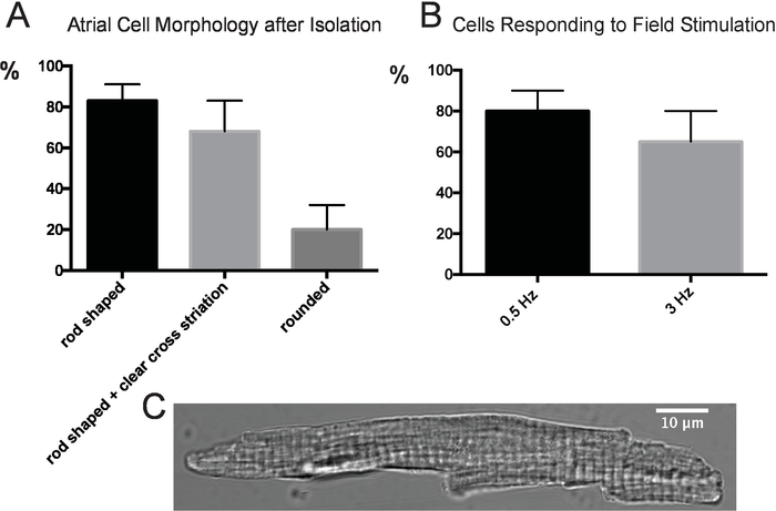
- Use a 10x objective to count all cells in the field of view and categorize them as rounded or rod-shaped. Determine how many of the rod-shaped cells show clear cross striation (switch to 25x objective if this is difficult to determine, see Figure 2C).
- Repeat steps 2.1–2.2.
- Calculate the percentages of rounded, rod-shaped and rod-shaped with clear cross striation cells.
- Modify the cell isolation procedure until 80% of cells are rod-shaped with clear cross-striation.
- Place 500 µL of the atrial cell suspension on a laminin-coated glass coverslip and place the cell chamber on the inverted microscope. Let the cells settle for 5 min. Perfuse with Tyrode’s solution containing 1.8 mM Ca2+.
- Use a cell stimulator system to start electrical field stimulation (2 ms bipolar pulse, 30 V) at 0.5 Hz and count the number of cells that contract in the field of view of a 10x objective.
- Repeat with external field stimulation at 3 Hz.
- Calculate the percentage of cells responding to 0.5 and 3 Hz stimulation rates (see Figure 2B). Refine cell isolation protocol until 50% of cells respond to 3 Hz field stimulation.
3. Na+ indicator loading of freshly isolated murine atrial myocytes
- Use the cell permeant acetoxymethyl (AM) ester of the fluorescent indicator sodium-binding benzofuran isophthalate (SBFI-AM).
- To facilitate dye dispersion and to achieve homogeneous cell loading dissolve SBFI in dimethyl sulfoxide (DMSO) and suitable surfactant polyols (e.g., pluronic) to achieve a final concentration of 10 μM SBFI in the cell suspension.
- Load the cells with SBFI for 60 min protected from light on a rocker at room temperature.
- Let the cells settle for 30 min, remove the supernatant and re-suspend the pellet in storage solution (1–2 mL).
NOTE: Experiments should be performed within 4 h of cell isolation. BDM is washed-out by perfusion with Tyrode’s solution prior to the start of experiments14.
4. Instrumentation, [Na+]i measurements and [Na+]i calibration
NOTE: Figure 3 depicts the light path schematic for the experimental instrumentation.
- Prepare calibration solutions with increasing Na+ concentrations as described in Tables 4–6.
- Add the permeabilizing agent gramicidin D (10 μM; from a stock solution stored at -20 °C) and the Na+, K+ ATPase inhibitor strophanthidin (100 μM, from a stock solution stored at -20 °C) to each calibration solution. Vortex well, or use a sonicator to ensure that gramicidin and strophantidin are completely dissolved.
- Fill a multi-barrel perfusion system with the calibration solutions containing the increasing [Na+]o prepared in steps 4.1 and 4.2. Connect to a cell chamber using slow perfusion rates (~1 mL/min; perfusion rates can vary with volume of the cell chamber).
- Connect suction to the cell chamber and collect perfusate in an appropriate (glass) container on the floor.
- Stop perfusion and suction.
- After allowing for 30 min of SBFI de-esterification, place 100 μL of the concentrated (step 3.5), dye-loaded cell suspension on a laminin-coated glass cover slip above the field of view of the inverted microscope’s 40x objective.
- Use a rapid switching illuminator with a 300 W xenon light source. Achieve wide field imaging with two excitation wavelengths (340 nm and 380 nm) using fast switching scanning mirrors and narrow bandwidth excitation filters (340 nm ± 10 nm; 380 nm ± 10 nm).
- Optimize the field of view of the EMCCD camera. A larger observation area requires longer frame times. Longer frame times lead to more bleaching of the indicator. Thus, balance the field of view size and frame time so that no noticeable bleaching of the probe occurs.
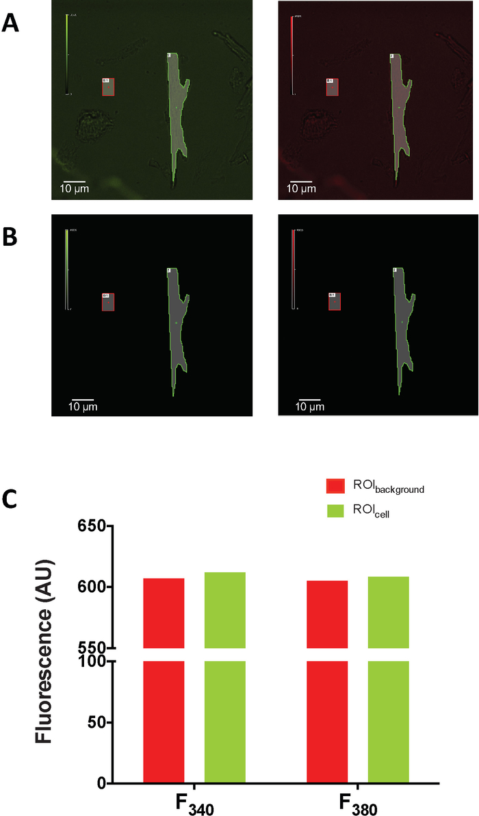
- Determine that there is only minimal intrinsic fluorescence in a subset of atrial cells that are not loaded with SBFI by ensuring that the cells’ intrinsic fluorescence is similar to background fluorescence (as shown in Figure 4).
- Determine the appropriate sampling rate depending on experimental design (sampling rates can be low because changes in [Na+]i are relatively slow (e.g. one data point acquired every 10–20 s).
- Attenuate the excitation light by using appropriate neutral density (ND) filters and by appropriately reducing the light source intensity (e.g., by altering intensity in the operating software).
- Collect the emission light at 510 ± 40 nm using appropriate filters with an EMCCD camera connected to the inverted microscope.
- Define a region of interest (ROI) for each cell and a background ROI (see Figure 4). Subtract the background from the recorded F340 and F380 signals (either online or during data analysis).
- Start the data acquisition.
- Restart perfusion and suction. Perfuse for 10–15 min with Tyrode’s solution containing 1.8 mM Ca2+ and record a stable F340/380 baseline before starting the experiment.
NOTE: Make sure to record a stable baseline for 10–15 min. If bleaching occurs (e.g., reduction of the F340/380 baseline) aim to further attenuate illumination intensity either by increasing ND filter density, decreasing excitation light signal intensity and/or acquisition frame time (see steps 4.10–4.11). - Start [Na+]i measurement according to research question.
5. [Na+]i Calibration
- After the conclusion of the experiment (step 4.16) perform a calibration of the F340/380 signal in each cell in situ.
- Calibrate the F340/380 signal by perfusing the SBFI-loaded myocytes with the calibration solutions (prepared in steps 4.1–4.3).
- Perfuse stepwise to elevate [Na+]o from 0 to 20 mM. Wait for stable F340/380 signal before moving to the next concentration (~5 min depending on flow rate; see Figure 3B and Figure 5A).
NOTE: The relation between the F340/380 signal and the [Na+] of the calibration solutions needs to be linear for a valid calibration of the F340/380 signal (Figure 5 and Figure 6).
Results
Evaluation of Atrial Cell Quality
Freshly isolated atrial myocytes were evaluated based on cell morphology and responsiveness to field stimulation as outlined in the protocol in six consecutive atrial cell isolations. Data shown in Figure 2 show a very high percentage of rod-shaped atrial myocytes that retain clear cross striation. Similarly, about 50% of atrial cells respond to high rates of external field stimulation up to 3 Hz.
SBFI Calibration
We evaluated the upper limit of [Na+]i that can be measured with SBFI in atrial myocytes by performing calibration experiments up to [Na+]o of 40 mM. We compared the linear regression of the SBFI calibration in one group of myocytes calibrated up to [Na+]o = 25 mM with calibration up to [Na+]o of 40 mM in a second group (n = 10 cells per experimental group). We found that calibrating atrial myocytes up to 40 mM [Na+]o significantly reduced the linearity of the calibration curve compared to the data obtained with calibration up to 25 mM [Na+]o. These data show that SBFI calibration is highly linear at 25 mM Na+ and that 40 mM Na+ is clearly outside the linear range of the indicator (Figure 6).
[Na+]i in Murine Atrial Myocytes
Murine atrial myocytes were freshly isolated and loaded with the Na+ selective fluorophore SBFI. The dye-loaded cells were seeded on a laminin coated glass cover slip and placed on an inverted microscope. The illumination light path is depicted in Figure 3. Cells are perfused with normal Tyrode. Figure 3B and Figure 5 depict typical experiments with subsequent calibration in situ. Figure 5A shows the F340/380 ratio during the experiment (Tyrode perfusion) followed by permeabilization of the cells with gramicidin and perfusion with solutions with 5 increasing [Na+]o. After perfusion with the highest [Na+]o cells are again perfused with 0 mM Na+ solution. Figure 5B shows the linear calibration curve, which is derived from the experiment depicted in Figure 5A. If the resulting calibration curve is non-linear, the experiment is not valid (as shown in Figure 6). Figure 5C shows the increase in the F340/380 ratio in the cell shown in Figure 5A during perfusion with increasing [Na+]o. Slow binding kinetics, variable degrees of dye compartmentalization and of de-esterification affect the signal to noise ratio and thus the sensitivity and precision of SBFI8,15,16. It is therefore important to perform a calibration of the F340/380 signal in situ for each cell in order to achieve reliable and precise measurements of [Na+]i.
[Na+]i in Atrial versus Ventricular Myocytes
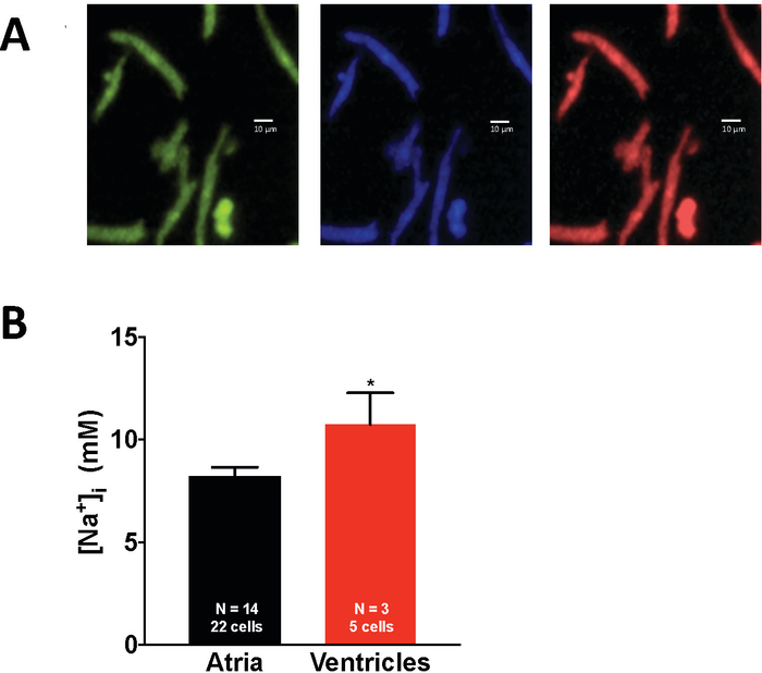
Although the objective of this protocol is to provide an optimization for the study of murine atrial myocyte [Na+]i, we also determined [Na+]i in a small subset of murine ventricular myocytes (Figure 7). Using this protocol we report [Na+]i in murine ventricular myocytes in line with previous results (10.74 ± 1.54 mM versus previously reported 11.1 ± 1.8 mM17 and 12 ± 1 mM18). Here we report the first quantitative measurements of [Na+]i in quiescent murine atrial myocytes (Figure 7). Similar to our previous findings in rabbit atrial myocytes1 [Na+]i in murine atrial myocytes is significantly lower than in ventricular myocytes, with a value of 8.17 ± 0.48 mM, which is ~30 % lower than in murine ventricular myocytes. To test for significance between groups the Student’s t-test was used. All data are shown as means ± SEM.

Figure 1: Isolation of murine atrial myocytes. (A) Retrograde cannulation of the mouse aorta. (B) Schematic depicting the positioning of the cannula well above the aortic sinuses. (C) View of the mouse heart during Langendorff perfusion and enzymatic digestion. (D) Isolated left and right mouse atrium after Langendorff perfusion. Please click here to view a larger version of this figure.

Figure 2: Atrial cell quality. (A) Percentage of rounded, rod-shaped and rod-shaped cells that retain clear cross striation after enzymatic isolation determined over six consecutive atrial cell isolations. (B) Percentage of atrial myocytes responding to different frequencies of external field stimulation determined over six consecutive atrial cell isolations. (C) Representative transmitted light image of atrial murine myocyte. Error bars denote SEM. Please click here to view a larger version of this figure.

Figure 3: Light path schematic and recording of SBFI fluorescence. (A) The illuminator used contains a full spectrum xenon lamp and fast switching mirrors that can direct the light to any of the available five excitation filter positions (here we use two positions for 340 nm and 380 nm). Emitted fluorescence (F340 and F380) is collected at 510 ± 40 nm by an EMCCD camera connected to an inverted microscope. (B) Recording of SBFI fluorescence and calibration in an atrial myocyte showing a stable baseline recording over an extended time period. Please click here to view a larger version of this figure.

Figure 4: Intrinsic fluorescence. (A) Example of typical white light images used for cell localization and ROI definition prior to data acquisition. The left panel shows white light images during UV illumination at 340 nm; the right panel at 380 nm in pseudocolor. (B) ROIs defined in (A) shown during UV illumination. (C) As a result of very low illumination intensity atrial intrinsic fluorescence is similar to background fluorescence. See text for further detail. Please click here to view a larger version of this figure.

Figure 5: Measurement of [Na+]i. (A) Representative experiment in an atrial myocyte during perfusion with Tyrode’s solution and subsequent in situ calibration using various [Na+]o solutions in the presence of 10 μM gramicidin D and 100 μM strophanthidin. (B) Fit of the calibration experiment depicted in (A) demonstrates high fidelity and a linear relationship between the F340/380 signal recorded during superfusion of permeabilized cells with increasing [Na+]o solutions. (C) Atrial myocyte F340/380 signal intensity increases during exposure to the calibration solutions of increasing [Na+]o depicted in (A). Please click here to view a larger version of this figure.

Figure 6: Linearity of SBFI calibration. (A) Example of an SBFI calibration up to 25 mM Na+. (B) Example of SBFI calibration up to 40 mM Na+. (C) Linear regression analysis of SBFI calibration with maximal [Na+]o of 25 mM versus 40 mM (10 cells per group). Please click here to view a larger version of this figure.

Figure 7: [Na+]i in quiescent atrial and ventricular myocytes. (A) From left to right, pseudocolor images from the simultaneous recording of emitted fluorescence at 340 nm excitation, 380 nm excitation and the fluorescence ratio (F340/380) in atrial myocytes loaded with SBFI. (C) [Na+]i in quiescent murine atrial and ventricular cardiac myocytes. *p < 0.05; error bars denote SEM; N = number of animals used. Please click here to view a larger version of this figure.
| CIB | Concentration (mM) |
| NaCl | 130 |
| KCI | 5.4 |
| MgCl2+6H2O | 0.5 |
| NaH2PO4 | 0.33 |
| Glucose | 16 |
| HEPES | 25 |
| Taurine | 6 |
| EGTA | 0-0.4* |
| CaCl2 | 0-0.15* |
| See text* |
Table 1. Cell isolation buffer. Solution used for initial perfusion of the heart. *See text for specific concentrations at each step.
| Enzymatic Solution | Composition |
| CIB | see Table 1 |
| Collagenase II | 0.8 mg/mL |
| Trypsin | 0.06 mg/mL |
| Protease XXIV | 0.06 mg/mL |
| CaCl2 | 0.1 mM |
Table 2. Enzyme Solution. This solution is used for the enzymatic digestion of the mouse heart. *See text for specific concentrations at each step.
| Modified Tyrode's Solution | Concentration (mM) |
| NaCl | 133 |
| KCl | 5 |
| MgCl2+6H2O | 2 |
| KH2PO4 | 1.2 |
| Taurine | 6 |
| Creatinine | 6 |
| Glucose | 10 |
| HEPES | 10 |
| 2,3-Butanedione monoxime (BDM) | 0 - 30* |
| Bovine Serum Albumin (BSA) | 0 - 15* |
| CaCl2 | 0 - 1.8* |
| see text* |
Table 3. Modified Tyrode's Solution. This solution is used for cell storage.
| Na+ Solution | Concentration (mM) |
| HEPES | 10 |
| Glucose | 10 |
| EGTA | 2 |
| NaCl | 30 |
| Na Gluconate | 115 |
| pH 7.2 with Trisbase |
Table 4. Na+ solution. This is a Na+ solution that is free of K+. It is used together with a K+ solution to achieve the different [Na+]o required for [Na+]i calibration.
| K+ Solution | Concentration (mM) |
| HEPES | 10 |
| Glucose | 10 |
| EGTA | 2 |
| KCl | 30 |
| K Gluconate | 115 |
| pH 7.2 with Trisbase |
Table 5. K+ solution. This is a K+ solution that is free of Na+. It is used together with the Na+ solution to achieve the different [Na+]o required for [Na+]i calibration.
| [Na+] (mM) | K+ (mM) | Na+ Solution (ml) | K+ Solution (ml) |
| 0 | 145 | 0 | 20 |
| 5 | 140 | 0.69 | 19.31 |
| 10 | 135 | 1.38 | 18.62 |
| 15 | 130 | 2.07 | 17.93 |
| 20 | 125 | 2.76 | 17.24 |
Table 6. Calibration solution. Schematic depicting the required volumes of the Na+ and K+ solutions to obtain the specific [Na+] required in the calibration step. The combined concentration of Na+ and K+ remains 145 mM at each [Na+].
Discussion
Here we introduce a novel EMCCD camera-based technique for the simultaneous quantitative measurement of [Na+]i in multiple viable atrial myocytes using sodium-binding benzofuran isophthalate (SBFI). The approach described here is the first to allow for the simultaneous measurement of [Na+]i in multiple cells. The main advantages this new protocol presenta are (i) the significant increase in experimental yield and (ii) the reduction in illumination intensity and duration due to the high sensitivity of the EMCCD camera, which significantly reduces cell damage.
Myocyte loading with the acetoxymethylester (AM) of SBFI takes significantly longer than cell loading with the AM of fluorescein-based chromophores (e.g., Fluo-3, Fluo-4; 45-90 min vs. 20 min, respectively). Additionally, SBFI-based quantitative measurements of [Na+]i, which require in situ calibration in each cell, are generally lengthy (60-90 min). Taken together, the time required for SBFI based quantitative [Na+]i measurements in single isolated cardiac myocytes limits experimental yield to 1 or 2 measurements per experimental day (in freshly isolated cells) before cell quality declines sharply. When using a traditional PMT-based method, only 1-2 cells can be measured per experimental day. This low experimental yield is problematic when the number of animals are limited (e.g., transgenic mice) or when differences in [Na+]i in the studied groups are expected to be small. Our novel approach allows for three to six (atrial) myocytes to be measured simultaneously, which significantly increases experimental yield.
Another important advantage our approach offers is the reduction of illumination intensity due to the use of an EMCCD camera. The high sensitivity of the camera allows for the use of significantly less illumination intensity. Additionally, the probe is only illuminated intermittently and not continuously, which further reduces light exposure. In addition, intrinsic fluorescence, which scales with the SBFI fluorescence is minimal as a result of the low illumination intensity and can be disregarded in this protocol.
Critical steps in the protocol and troubleshooting
Dye-loading: Previous protocols using cell permeant AM forms of SBFI to load cardiac myocytes were performed at 37 °C1. The fragile murine atrial myocytes were affected by high temperature and atrial myocyte quality suffered significantly during 37 °C dye loading. In this protocol we optimized SBFI dye loading at room temperature by using a combination of a specific mix of surfactant polyols in addition to DMSO and mechanic agitation (cell rocker). The switch from 37 °C to room temperature during dye loading led to a significant increase in cell quality, which was assessed as the percentage of cells responding to external field stimulation.
Acquisition parameters: Changes in [Na+]i are relatively slow (seconds) compared to changes in [Ca2+]i and acquisition does not require high temporal resolution. Therefore, acquisition sampling frequency can be relatively low.
Intrinsic fluorescence: Due to the very low illumination intensity used in our system autofluorescence was similar to background fluorescence (Figure 4). This needs to be verified for each illumination setup and after changes in illumination intensity are implemented. If intrinsic fluorescence is significant it should be subtracted as previously described1,11.
Optimization of the illumination pathway: While SBFI is the only Na+-sensitive fluorophore that allows for ratiometric Na+ quantification due to a true spectral shift upon Na+ binding, the increase in fluorescence is low when compared to fluorescein-based indicators. It is therefore important to optimize the optical pathway to prevent bleaching. This is best achieved by testing different neutral density filters. It is imperative to have a stable baseline of F340/380 over at least 10 min before starting the experiment. As changes in F340/380 are small and experiments are fairly long, the lack of a stable baseline will result in significant errors in calibration.
Alternative methods for quantitative measurements of [Na+]i in living cells
Na+ sensitive microelectrodes
There are other techniques to measure [Na+]i, most importantly by using Na+-sensitive microelectrodes introduced by Hinke9,19 and modified by Thomas20. Na+-sensitive microelectrodes are most suitable for [Na+]i measurements in large cells like snail neurons21, amphibian pregastrular embryos22 and cardiac Purkinje fibers23. Because this technique requires the penetration of one or two glass electrode tips into the cytoplasm, cell integrity in smaller cells, like atrial myocytes which are narrow, is easily compromised. Moreover, this technique can lead to leakage of extracellular Na+ or electrode solution into the cytoplasm. This effect is aggravated in cells with small cytosolic volumes11. Fluorescence indicators are, therefore, preferable for measuring Na+ in small and narrow cells like atrial myocytes or sino-atrial nodal cells.
[Na+]i measurements using photomultiplier tubes
Photomultiplier tube (PMT)-based fluorescence measurements of [Na+]i represent the majority of [Na+]i measurements in isolated cardiac myocytes. The biggest limitation of PMT-based acquisition of [Na+]i is the restriction of measurements to one cell at a time. Due to the length of quantitative measurements of [Na+]i using SBFI this results in only one or two cells measured per experimental day. In fact, this restriction of PMT-based SBFI measurements motivated the exploration of a camera based approach, which allows for the simultaneous acquisition of multiple cells. In addition, while providing excellent temporal resolution, there is no spatial resolution of the whole cell fluorescence signal recorded by the PMT. Compared with EMCCD cameras, PMT sensitivity is less and thus requires a higher illumination intensity, which results in a higher probability of significant bleaching and reactive oxygen species-induced alteration of cell physiology.
[Na+]i measurements using two-photon excitation microscopy
Despa et al. measured [Na+]i in ventricular myocytes using two-photon microscopy24. This approach provides excellent spatial resolution which the authors used to demonstrate intracellular [Na+]i gradients induced by local inhibition of the Na+/K+-ATPase. Using two-photon microscopy for measurements of [Na+]i requires an in vivo determination of SBFI Kd as well as prior determination of single-photon excitation spectra of SBFI with various [Na+]24. Because of a high degree of complexity required for two-photon microscopy this technique is best suited to research questions that require a high degree of spatial resolution and quantitative measurements of [Na+]i. A slightly lesser degree of spatial resolution can be achieved with the camera-based technique, which is less complex and allows for simultaneous acquisition in multiple cells.
Single-excitation fluorescent measurements of [Na+]i
Kornyeyev et al. have used the single wavelength Na+ indicator Asante NaTRIUM Green-2 (ANG) to perform quantitative measurements of [Na+]i in rabbit ventricular myocytes25. Single wavelength indicators are generally favored for qualitative measurements due to their large dynamic range. Indicator calibration is less straightforward than in dual wavelength indicators, where the ratio of two fluorescence signals peaking at different wavelength is used to minimize dye concentration and distribution and wavelength-independent variations in illumination intensity and detection efficiency. ANG’s discrimination against K+ is poor. In fact, in a cardiac myocyte the majority of ANG fluorescence is due to its K+ binding. Additionally, another study determined ANG Kd in cells as 56 mM Na+, which is well above the physiological [Na+]i in cardiac myocytes. Sodium Green is a single wavelength Na+ indicator with better discrimination against K+ than SBFI (41 fold) and might be a better candidate for quantitative measurements of [Na+]i if the use of a single wavelength indicator is desirable26.
Variations in indicator loading, acquisition and calibration for use with camera based [Na+]i measurements
Pipette based loading of SBFI
The tetra-ammmonium salt of SBFI7, which is water soluble, can be delivered into the cytosol directly through a patch pipette. The advantage of using an SBFI salt is a significant shortening of the loading time. In addition, if the kd of the salt is experimentally determined, calibration curves can be recorded in a few cells and then used in further experiments. This eliminates the need for calibration in each cell. The disadvantage of this method is the loss of cell integrity and intracellular milieu, which will be equilibrated with the contents of the pipette solution. Cellular electrophysiology techniques (voltage clamp technique in whole cell configuration) are required as compromised cell integrity necessitates an externally applied (‘clamped’) membrane potential. Moreover, most pipette solutions require the addition of Ca2+ chelators (e.g. ethylene glycol-bis(β-aminoethyl ether)-N,N,N′,N′-tetraacetic acid [EGTA]), which can interfere with and even suppress the intracellular Ca2+ transient. Thus, when Na+ measurements are performed in conjunction with evaluation of intracellular Ca2+ homeostasis maintaining cell integrity may be desirable. In addition, simultaneous measurements in multiple cells cannot be performed using pipette-based dye loading.
Null point calibration using SBFI-AM
Use of SBFI-AM, as we show here, has the advantage of being loaded into the cell without compromising cell integrity. The acetoxymethylester is cell membrane permeable. Ester groups are cleaved by cytosolic esterases leaving the indicator in the cytosol10. A modification of the calibration technique used in this protocol is the null-point approach, which was developed by Despa and colleagues11. After completion of [Na+]i measurements the cell is permeabilized and perfused with solutions containing different [Na+]o in the range of the anticipated values for [Na+]i. When the perfusion with a specific [Na+]o results in no further change in the F340/380 ratio [Na+]i = [Na+]o. This approach offers a faster calibration than described in this protocol. However, it presents an important limitation for the atrial cell studies proposed here. The null point method can only assess the [Na+]i directly before permeabilization has started. Any measurements of [Na+]i that lead to changes in [Na+]i during the experiment (e.g., a range of frequency dependent changes in [Na+]i) cannot be reliably calibrated.
[Na+]i measurements with SBFI in dual emission mode
Baartscheer et al. showed that single excitation of SBFI-loaded rat ventricular cells at 340 nm leads to an exclusively sodium-dependent fluorescence emission in the range 400-420 nm and very little change in fluorescence above 530 nm upon replacement of sodium by potassium16. These spectral and quantum efficiency changes allow SBFI excited at 340 nm to be used in a dual emission ratio mode (410 nm and 590 nm).
Comparison with previous work
Here, we determined [Na+]i in murine ventricular myocytes using a camera based approach and found our results in line with previous studies17,18. The atrial data acquired using this protocol represent the first quantitative measurements of [Na+]i in viable murine atrial myocytes. In line with our previous findings in rabbit atrial myocytes1 [Na+]i in murine atrial myocytes is lower than in murine ventricular myocytes (Figure 5).
Future Outlook
Using a camera-based approach for the measurement of [Na+]i offers the possibility of evaluating different cellular compartments (e.g., nucleus, cytosol) because the camera offers the advantage of higher spatial resolution compared with PMT or electrode based measurements without the degree of complexity required for two-photon microscopy.
In summary, the method presented here allows for the first time to measure [Na+]i reliably simultaneously in multiple atrial myocytes. As demonstrated, the EMCCD camera has a higher sensitivity than state of the art PMTs and requires only very low illumination intensity. This leads to the elimination of significant cellular intrinsic florescence in our optical setup. This technique can be used to study the effect of different stimulation rates and various drug effects on [Na+]i, and since multiple measurements can be performed simultaneously, the power to detect differences is augmented significantly.
Disclosures
The authors have nothing to disclose.
Acknowledgements
This work was supported by a Scientist Development Grant from the American Heart Association (14SDG20110054) to MG; the NIH Interdisciplinary Training Grant in Muscle Biology (T32 AR007592) and the NIH Cardiovascular Disease Training Grant (2T32HL007698-22A1) to LG; a Scientist Development Grant from the American Heart Association (15SDG22100002) to LB and by NIH grants R01 HL106056, R01 HL105239 and U01 HL116321 to WJL.
Materials
| Name | Company | Catalog Number | Comments |
| 2,3-Butanedione monoxime (BDM) | Sigma-Aldrich | B0753 | |
| 340 Excitation Filter | Chroma | ET40X | 25 mm |
| 380 Excitation Filter | Chroma | ET80X | 25 mm |
| 510 Emission Filter | Chroma | ET510/80m | 25 mm |
| Bovine Serum Albumin (BSA) | Sigma-Aldrich | A7906 | |
| Bubble trap | BD Medical Technologies | 904477 | Custom made from a 5 ml Luer Lok Syringe, which is located in the tubing path from the perfusing solution to the cannula |
| CaCl2 solution | Sigma-Aldrich | 21115 | |
| Cannula | BD Medical Technologies | 305167 | Custom made from a 22 G x 1 1/2 inch needle. Cut to 1 inch and sand 1mm distal tip. |
| Cell Chamber | Custom machined with an opening that can securely hold a 25 mm glass cover slip and with a cover that has an inlet and an outlet port for perfusion. | ||
| Circulating Water Bath | VWR | ||
| Collagenase II | Worthington | LS004176 | Specific activity 290 U/g |
| Creatinine | Sigma-Aldrich | C0780 | |
| DG5-plus illuminator | Sutter Instrument | Lambda DG-4/DG-5 Plus | |
| DMSO | Thermo Fischer | BP231 | |
| EGTA | Sigma-Aldrich | E4378 | |
| EMCCD camera | Princeton Instruments | ProEM-HS | |
| Fine Hemostats | Fine Science Tools | 130-20 | |
| Fine Scissors | Fine Science Tools | 14060-10 | |
| Forceps Supergrip | Fine Science Tools | 00632-11 | |
| Glass Cover slips | VWR | 4838089 | 25 mm circle |
| Glucose | Sigma-Aldrich | G7528 | |
| Gramicidin D | Sigma-Aldrich | G5002 | |
| HEPES | Sigma-Aldrich | H3375 | |
| Inner silicon Tubing | VWR | VWRselect brand silicon tubing | |
| Inverted microscope | Nikon Instruments | NikonTE 2000 U | |
| Isolation Tools | |||
| K Gluconate | Sigma-Aldrich | P1847 | |
| KCI | Sigma-Aldrich | P5405 | |
| KH2PO4 | Calbiochem | 529568 | |
| Langendorff perfusion apparatus | |||
| MgCl2.6H2O * | Sigma-Aldrich | M0250 | |
| MyoPacer Cell Stimulator | IonOptix | ||
| Na Gluconate | Sigma-Aldrich | S2054 | |
| NaCl | Sigma-Aldrich | S9888 | |
| NaH2PO4 | Sigma-Aldrich | S9390 | |
| Natural Mouse Laminin | Thermo Fischer | 23017015 | 0.5-2.0 mg/ml |
| Outer tubing | VWR | ||
| Petri dish 35X10 mm | Falcon | 351008 | |
| PowerLoad | Thermo Fischer | P10020 | |
| Protease XXIV | Sigma-Aldrich | P8038 | |
| SBFI-AM | Thermo Fischer | S1264 | |
| Silk suture | Fine Science Tools | 18020-50 | 0.12 mm diameter |
| Small Spring scissors | Fine Science Tools | 15000-03 | |
| Standard Pattern Forceps | Fine Science Tools | 11000-12 | |
| Strophanthidin | Sigma-Aldrich | G5884 | |
| Surgical Scissors Tough Cut | Fine Science Tools | 14054-13 | |
| Suture Tying Forceps | Fine Science Tools | 00272-13 | |
| Taurine | Sigma-Aldrich | T0625 | |
| Trisbase | Sigma-Aldrich | TRIS-RO | |
| Trypsin | Sigma-Aldrich | T0303 | |
| UVFS Reflective 0.1 ND Filter | Thorlabs | NDUV01B | 25 mm |
| UVFS Reflective 0.2 ND Filter | Thorlabs | NDUV02B | 25 mm |
| UVFS Reflective 0.3 ND Filter | Thorlabs | NDUV03B | 25 mm |
| UVFS Reflective 0.5 ND Filter | Thorlabs | NDUV05B | 25 mm |
| UVFS Reflective 1 ND Filter | Thorlabs | NDUV010B | 25 mm |
References
- Greiser, M., et al. Tachycardia-induced silencing of subcellular Ca2+ signaling in atrial myocytes. Journal of Clinical Investigation. , (2014).
- Greiser, M., Lederer, W. J., Schotten, U. Alterations of atrial Ca(2+) handling as cause and consequence of atrial fibrillation. Cardiovascular Research. 89, 722-733 (2011).
- Voigt, N., et al. Enhanced sarcoplasmic reticulum Ca2+ leak and increased Na+-Ca2+ exchanger function underlie delayed afterdepolarizations in patients with chronic atrial fibrillation. Circulation. 125, 2059-2070 (2012).
- Bers, D. M. Cardiac excitation-contraction coupling. Nature. 415, 198-205 (2002).
- Sossalla, S., et al. Altered Na(+) currents in atrial fibrillation effects of ranolazine on arrhythmias and contractility in human atrial myocardium. Journal American College of Cardiology. 55, 2330-2342 (2010).
- Wan, E., et al. Aberrant sodium influx causes cardiomyopathy and atrial fibrillation in mice. Journal of Clinical Investigation. 126, 112-122 (2016).
- Minta, A., Tsien, R. Y. Fluorescent indicators for cytosolic sodium. Journal of Biological Chemistry. 264, 19449-19457 (1989).
- Donoso, P., Mill, J. G., O'Neill, S. C., Eisner, D. A. Fluorescence measurements of cytoplasmic and mitochondrial sodium concentration in rat ventricular myocytes. Journal of Physiology. 448, 493-509 (1992).
- Friedman, S. M., Jamieson, J. D., Hinke, J. A., Friedman, C. L. Use of glass electrode for measuring sodium in biological systems. Proceedings of the Society for Experimental Biology. 99, 727-730 (1958).
- Harootunian, A. T., Kao, J. P., Eckert, B. K., Tsien, R. Y. Fluorescence ratio imaging of cytosolic free Na+ in individual fibroblasts and lymphocytes. Journal of Biological Chemistry. 264, 19458-19467 (1989).
- Despa, S., Islam, M. A., Pogwizd, S. M., Bers, D. M. Intracellular [Na+] and Na+ pump rate in rat and rabbit ventricular myocytes. Journal of Physiology. 539, 133-143 (2002).
- Shioya, T. A simple technique for isolating healthy heart cells from mouse models. Journal of Phsiological Sciences. 57, 327-335 (2007).
- Icardo, J. M., Colvee, E. Origin and course of the coronary arteries in normal mice and in iv/iv mice. Journal of Anatomy. 199, 473-482 (2001).
- Yu, Z. B., Gao, F. Non-specific effect of myosin inhibitor BDM on skeletal muscle contractile function]. Zhongguo Ying Yong Sheng Li Xue Za Zhi. 21, 449-452 (2005).
- Levi, A. J., Lee, C. O., Brooksby, P. Properties of the fluorescent sodium indicator "SBFI" in rat and rabbit cardiac myocytes. Journal of Cardiovasc Electrophysiology. 5, 241-257 (1994).
- Baartscheer, A., Schumacher, C. A., Fiolet, J. W. Small changes of cytosolic sodium in rat ventricular myocytes measured with SBFI in emission ratio mode. Journal of Molecular and Cellular Cardiology. 29, 3375-3383 (1997).
- Despa, S., Tucker, A. L., Bers, D. M. Phospholemman-mediated activation of Na/K-ATPase limits [Na]i and inotropic state during beta-adrenergic stimulation in mouse ventricular myocytes. Circulation. 117, 1849-1855 (2008).
- Correll, R. N., et al. Overexpression of the Na+/K+ ATPase alpha2 but not alpha1 isoform attenuates pathological cardiac hypertrophy and remodeling. Circulation Research. 114, 249-256 (2014).
- Hinke, J. Glass micro-electrodes for measuring intracellular activities of sodium and potassium. Nature. 184 (Suppl 16), 1257-1258 (1959).
- Thomas, R. C. New design for sodium-sensitive glass micro-electrode. Journal of Physiology. 210, 82P-83P (1970).
- Thomas, R. C. Membrane current and intracellular sodium changes in a snail neurone during extrusion of injected sodium. Journal of Physiology. 201, 495-514 (1969).
- Slack, C., Warner, A. E., Warren, R. L. The distribution of sodium and potassium in amphibian embryos during early development. Journal of Physiology. 232, 297-312 (1973).
- Eisner, D. A., Lederer, W. J., Vaughan-Jones, R. D. The control of tonic tension by membrane potential and intracellular sodium activity in the sheep cardiac Purkinje fibre. Journal of Physiology. 335, 723-743 (1983).
- Despa, S., Kockskamper, J., Blatter, L. A., Bers, D. M. Na/K pump-induced [Na](i) gradients in rat ventricular myocytes measured with two-photon microscopy. Biophysical Journal. 87, 1360-1368 (2004).
- Kornyeyev, D., et al. Contribution of the late sodium current to intracellular sodium and calcium overload in rabbit ventricular myocytes treated by anemone toxin. American Journal of Physiology-Heart and Circulatory Physiology. 310, H426-H435 (2016).
- Szmacinski, H., Lakowicz, J. R. Sodium Green as a potential probe for intracellular sodium imaging based on fluorescence lifetime. Annals of Biochemistry. 250, 131-138 (1997).
Reprints and Permissions
Request permission to reuse the text or figures of this JoVE article
Request PermissionThis article has been published
Video Coming Soon
Copyright © 2025 MyJoVE Corporation. All rights reserved