Method Article
An Experimental Model of Myocardial Infarction for Studying Cardiac Repair and Remodeling in Knockout Mice
* These authors contributed equally
In This Article
Summary
Here, we describe an experimental model of myocardial infarction, an echocardiography procedure to study cardiac remodeling and function, and procedures for quantifying fibrosis and hypertrophy in picrosirius red-stained and rhodamine-stained sections, as well as the infarct size and expansion index in slices stained with Masson's trichrome.
Abstract
Cardiovascular disease is the most prevalent cause of death in Western countries, with acute myocardial infarction (MI) being the most prevalent form. This paper describes a protocol for studying the role of galectin 3 (Gal-3) in the temporal evolution of cardiac healing and remodeling in an experimental animal model of MI.
The procedures described include an experimental model of MI with a permanent coronary ligature in male C57BL/6J (control) and Gal-3 knockout (KO) mice, an echocardiography procedure to study cardiac remodeling and systolic function in vivo, a histological evaluation of interstitial myocardial fibrosis with picrosirius red-stained and rhodamine-conjugated lectin-stained sections for studying myocyte hypertrophy by the cross-sectional area (MCSA), and the quantification of infarct size and cardiac remodeling (scar thinning, septum thickness, and expansion index) by planimetry in slices stained with Masson's trichrome and triphenyl tetrazolium chloride. Gal-3 KO mice with MI showed disrupted cardiac remodeling and an increase in the scar thinning ratio and the expansion index. At the onset of MI, myocardial function and cardiac remodeling were also severely affected. At 4 weeks post MI, the natural evolution of fibrosis in infarcted Gal-3 KO mice was also affected.
In summary, the experimental model of MI is a suitable model for studying the temporal evolution of cardiac repair and remodeling in mice with the genetic deletion of Gal-3 and other animal models. The lack of Gal-3 affects the dynamics of cardiac repair and disrupts the evolution of cardiac remodeling and function after MI.
Introduction
Myocardial infarction (MI) is the most prevalent form of cardiovascular disease. After MI, the myocardium undergoes serial morphological and functional changes, including the healing of the MI infarct zone, ventricular remodeling (VR), and myocardial dysfunction1. The healing of MI is a dynamic and well-orchestrated process associated with profound inflammatory infiltration that ends in the formation of a fibrotic scar2,3. The experimental model of MI in mice is currently used for studying cardiac remodeling under pathological conditions4,5, and awareness of the precise surgical protocol is essential to develop a reproducible and effective procedure for inducing a permanent coronary ligature. This method is needed to study the healing of MI and its relevance in the temporal evolution of left ventricular remodeling (LVR) and the cardiac dysfunction associated with MI.
Galectins are a group of lectins that recognize specific carbohydrates in intracellular ligands, membrane receptors, and extracellular glycoproteins. Galectin 3 (Gal-3) is a member of this family that acts through the recognition and cross-linking of N- and O-glycans in glycoconjugates on the cell surface, and it is widely expressed in the immune system6. Previous studies have investigated the role of Gal-3 as a regulator of inflammation and fibrosis in cardiovascular diseases7,8,9,10,11,12. As targeting the regulatory factors of inflammation during healing is highly relevant because inflammation can notably affect the evolution of remodeling, we aimed to describe a protocol for studying the temporal evolution of post-MI ventricular remodeling and the steps and methods for determining how the genetic mutation of Gal-3 modifies the temporal evolution of healing in MI and affects cardiac remodeling and function in mice.
Protocol
NOTE: All the experiments described in this protocol were approved by the Animal Care and Research Committee of the University of Buenos Aires (CICUAL), in line with the National Research Council (US) Committee for the Update of the Guide for the Care and Use of Laboratory Animals13. For the experiments, use male, age-matched C57BL/6J and Gal-3 KO mice (8-10 weeks old) weighing 30-35 g, which allow for better manipulation for surgery. Allow the animals access to water and food ad libitum. The Gal-3 KO mice were bred on a C57BL/6J background at the same bioresource facilities as the control C57BL/6J mice.
1. Surgical area and instruments
- Before starting the surgery, verify that the ventilator is plugged into a power source and working correctly. Confirm that there is enough anesthetic solution. Prepare the solution on the day of the surgery by calculating the number of animals to be used.
- Confirm that all the surgical instruments are sterilized, including stainless scissors, micro scissors, needle holders for big and small needles, retractors, extra sharp, curved forceps, serrated forceps, and tissue forceps. Clean the working area with 70% ethanol.
- Ensure the availability of small cotton balls, hyssops, and gauze for immediate cauterization of any potential hemorrhage.
- Keep ready-prepared 10-0 to 7-0 silicone-coated, braided silk sutures for ligature of the left descending coronary artery (LDA), nylon suture for thorax closing, and linen thread for closing the skin.
2. Anesthesia and intubation
- Weigh the mice to determine the dose of anesthesia.
- Prewarm a heating pad surrounded by a Styrofoam base to 40 °C.
- Anesthetize the mice with intramuscular administration of 0.1 mL/10 g of body weight of a solution containing ketamine (65 mg/kg), xylazine (13 mg/kg), and acepromazine (1.5 mg/kg).
- Once the mouse is anesthetized and before starting the surgical procedure, check the anesthetic depth by inducing a slightly painful stimulus, such as by pressing the feet with the fingers. If the animal responds to the stimulus, adjust the anesthetic depth.
- Place the mouse in dorsal decubitus, and tape its legs to the working base above the heating pad, placing it under a binocular stereo microscope. Hyperextend the neck with a thread holding the maxillary teeth attached to the working base.
- Then, for the intubation part, expose the trachea rings through a minimal incision in the neck, and channel through it with a 20 G intravenous catheter connected to a rodent ventilator (tidal volume: 250 mL/stroke) at a respiratory frequency of 34-38 cycles/min, as described previously14.
- Place the animal in lateral decubitus to perform the left lateral thoracotomy at the fourth or fifth intercostal space. Make an incision in the animal's skin; observe the muscles below, and spread them carefully to separate them from the thoracic wall to clearly see the intercostal space. At this point, ensure that the animal is properly connected to the ventilator; then, open the intercostal space by making a hole with the extra sharp forceps.
NOTE: The LDA should be identifiable along the free left ventricular (LV) wall in the intercostal spaces mentioned above. - Perform the pericardiectomy, and identify the LDA by contrasting the coronary arteries with the coronary veins and the LDA ramification below the left auricle. Then, perform the ligature of the LDA using the 8-0 silk thread ~2 mm from the edge of the auricle. Finally, close the thorax by layers using the 6-0 silk thread-close the ribs, ensuring that there is no pneumothorax inside (do this carefully by forcing the lungs to expand with the ventilator), and approach the muscles or suture them before closing the skin.
- Once the skin is closed, slowly disconnect the mouse from the ventilator in ventral decubitus, and remove the endotracheal tube when the respiratory frequency has recovered. Let the animal recover from the anesthesia in a quiet environment and, preferably, with a stable room temperature at 27 °C.
- Wait for the animals to recover from the anesthesia, begin moving their limbs, and for their normal respiratory frequency to be restored. Then, house them in individual cages until the end of the protocol.
- Perform the same procedure on the control or sham-operated animals but without the ligature of the LDA.
3. Study design
- For testing the temporal evolution of healing and ventricular remodeling after MI, randomize the mice into the following groups:
- To study the early phase of ventricular remodeling after 1 week of MI evolution, assign the mice to the following groups: 1) C57 Sham (1 week); 2) Gal-3 KO Sham (1 week); 3) C57 MI (1 week); 4) Gal-3 KO MI (1 week).
- To study the late phase of ventricular remodeling at 4 weeks of MI, assign the mice to the following groups: 5) C57 Sham (4 weeks); 6) Gal-3 KO Sham (4 weeks); 7) C57 MI (4 weeks); 8) Gal-3 KO MI (4 weeks).
4. Echocardiography
NOTE: For mouse echocardiograms, linear transducers over 10 MHz must be used for proper visualization of the wall diameters and cavity sizes. This procedure can be performed under anesthesia with intraperitoneal (IP) avertin at 1.15 mL/kg or in conscious animals. However, the latter can lead to confounding results in mice with MI due to the stress and anxiety caused by manipulation.
- To anesthetize a mouse, pick it up, hold it with its back toward the palm, and turn it over to reach the surface of the abdomen. In that position, inject the IP anesthesia at a 45° angle between the animal and the subcutaneous needle.
- Once the mouse is anesthetized, shave its chest, and place the mouse over a prewarmed heating pad in the dorsal decubitus position. To obtain parasternal long-axis and short-axis views, move the transducer 90°. Once the correct axis view is obtained, place the cursor at the papillary muscle level, press the 2-D M-mode key to capture the images, and use image analysis software to measure the following parameters:
- Measure the LV dimensions, including the wall thickness (LVWT), the LV areas both in systole (S) and diastole (D), and the left ventricular diastolic area (LVDA) and left ventricular systolic area (LVSA) in at least three beats.
- In addition, calculate the ventricular function by the ejection fraction (EF, %), the shortening fraction (SF, %), and the cardiac mass (assume an uncorrected cube) using equation (1), equation (2), and equation (3), as previously described15.
SF (%) = ([LVEDD - LVESD]/LVEDD) × 100 (1)
EF (%) = ([LVDA - LVSA]/LVDA) × 100 (2)
LV mass = 1.055 × ([IVST + LVEDD + PWT]3− [LVEDD]3) (3)
Where LVEDD is the left ventricular end-diastolic diameter, LVESD is the left ventricular end-systolic diameter, IVST is the intraventricular thickness, and PWT is the posterior wall thickness.
5. Histology evaluation
- During necropsy, extract the heart from the animal by opening the thorax from side to side and cutting all the structures that surround it. Clean the blood clot that is inside the cavities by applying gentle pressure using tissue paper.
- Harvest and weigh the heart on a laboratory precision scale. Immerse it in 10% formaldehyde for at least 72 h at room temperature. Cut the heart manually from the apex to the base in 1 mm thick transverse slices using a blade, and process the slices by embedding them in paraffin. Make serial cuts to the paraffin-embedded sections of 5 µm thickness with a microtome.
- Place each section between slides, and stain them with hematoxylin and eosin (H&E), Masson's trichrome, rhodamine-conjugated lectin-stained sections, or Picrosirius red15,16.
- By using an appropriate photomicroscope, take digitalized images at 400x for morphometry, fibrosis, and MCSA quantification. Verify that the microscope is attached to a digital camera and connected to a computer with image analysis software. For each morphometric analysis, ensure that the images are in the same areas, with a minimum of 10 high power fields at 400x per section (septum, infarcted zone, and remote zone) and without overlapping the fields.
- To measure the MCSA, be aware of the positions of the cardiac myocytes, and count only those myocytes that are transversally sliced and surrounded by at least three nearby capillaries.
- In the Picrosirius red-stained slices, identify the scar and septum areas, and image the interstitial collagen in both zones. Upload the images onto the analysis software, and open the threshold tab to highlight all the positive and negative collagen areas To obtain the data, press the measure tab, and save the results. For the calculation of the percentage of collagen per region, use equation (4), and add the collagen-positive zones and divide them by the total tissue, including the collagen-positive areas, as described elsewhere15,16.
Collagen (%) = Picrosirius red area/Total tissue area (4) - Quantify the MCSA from the digitalized images obtained from the rhodamine-conjugated lectin-stained sections of the paraffin-embedded samples. To obtain the correct image, use image analysis software to trace the red outlines of the myocytes surrounding the cell membranes. Select the area tab, trace the outlines, and press the measure tab function. Finally, save the results from the cell areas16.
- By using an appropriate photomicroscope, take digitalized images at 400x for morphometry, fibrosis, and MCSA quantification. Verify that the microscope is attached to a digital camera and connected to a computer with image analysis software. For each morphometric analysis, ensure that the images are in the same areas, with a minimum of 10 high power fields at 400x per section (septum, infarcted zone, and remote zone) and without overlapping the fields.
6. Quantitative determination of infarct size and planimetry to evaluate cardiac remodeling
- Measure the myocardial infarct size, the wall thickness, and the length of the endocardial and epicardial circumferences using planimetry from the histological images of the Masson's trichrome-stained sections obtained with a light microscope (4x) and the appropriate software.
- For the quantification of the infarct size, identify the infarct zone (blue) and the remote zone (red). Trace and measure the total length of the infarct zone and the remote zone at the endocardial and epicardial sides. Calculate the average of the endocardial and epicardial tracings as a percentage of the total LV circumference17.
- Similarly, measure the scar thickness (the average of five equidistant measurements) and the septum thickness (the average of three equidistant measurements) in a middle section of the heart, and use these measurements to determine the scar thickness ratio (equation [5]) and the expansion index (equation [6]18).
NOTE: All the values can be recorded in a spreadsheet.
Scar thickness ratio = Scar thickness/Septum thickness (5)
Expansion index = (LV cavity area/total LV area) × (septum thickness/scar thickness) (6)
NOTE: Since the remodeling may modify the expansion, resulting in underestimation or overestimation of the infarct size, some time may be needed to perform a pilot experiment for measuring the infarct size at 24 h, followed by euthanasia. In this case, anesthetize the animal with the same IP anesthetic solution used for the MI procedure.
- Place the animal in dorsal decubitus, and intubate it as described earlier. Once the animal is anesthetized, make a deep diagonal incision that reaches the skin, muscles, and costal bones from the xiphoid apophysis to the axillary hollow.
- Isolate the aortic arch, make a small hole in ascending aorta, and introduce a catheter to perfuse the heart with Evan's blue. Then, manually remove the stained heart from the animal, and cut it from the apex to the base with a sharp blade. Place the heart slices in 1% triphenyl tetrazolium chloride (TTC) in isotonic phosphate buffer (pH 7.4) and incubate at 37 °C for 30 min 4 to confirm that the animals are comparable in terms of infarct size.
Results
Post-MI survival and necropsy
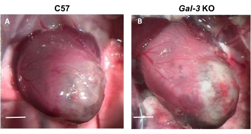
Over 4 weeks of follow-up, 17% (4/23) of the C57 mice versus 40% (8/20) of the Gal-3 KO mice were found dead. The necropsy was performed; the dead Gal-3 KO mice showed larger hearts than the C57 mice (Figure 1), and 38% of the C57 mice compared with 32% of the Gal-3 KO mice had macroscopic chest clots that were directly associated with cardiac rupture. The latter demonstrated that the cause of death was not related to cardiac rupture. By the end of the protocol and after the necropsy, cardiac remodeling could be identified by simple macroscopic visualization of the heart's necrotic areas. Figure 1 shows a representative image of the hearts of both experimental groups, in which an increase in the size of the hearts, the extension, and the presence of aneurism of the scar was primary identified and found to be markedly increased in the hearts of Gal-3 KO mice with MI.
Cardiac remodeling and function in vivo
The first approach for evaluating cardiac remodeling and function in vivo is echocardiography, which is a non-invasive method to assess cardiac geometry and function. Thus, echocardiography can be used to quantify LV dilation, LV wall thickness, and contractility at different time points of MI. Figure 2B,C show representative echocardiograms of Gal-3 KO mice (Figure 2B) and C57 mice (Figure 2C) at 1 week post MI. Interestingly, the Gal-3 KO mice showed larger LV cavity size than the C57 group (Figure 2B,C), showing that the deficiency of Gal-3 had a deleterious effect on cardiac remodeling and myocardial function after MI. To estimate systolic function, the quantification of morphological parameters is needed. In these experimental conditions, chronic LV function was significantly reduced in both genotypes with MI.
Histology and morphometry
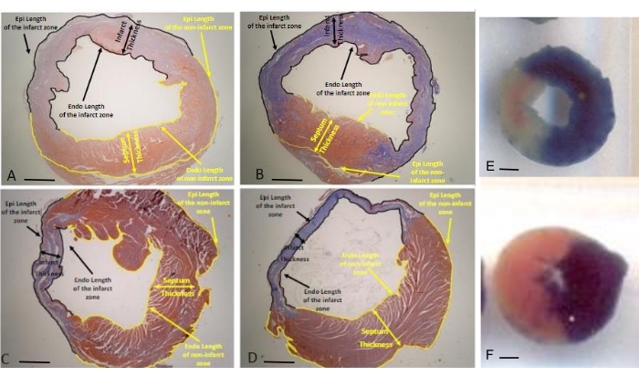
Cardiac remodeling can be analyzed by quantifying the infarct size and morphometric parameters in slices stained with Masson's trichrome by quantitative planimetry. With this stain, the infarct zone is stained blue, and the healthy cardiac tissue is stained slightly red. Once these colors are identified, the respective areas are carefully determined by drawing a perimeter line and measuring them using image analysis software (Figure 3).
Expansion is one of the first phenomena to occur after post-MI remodeling. The remodeling can lead to the overestimation of the infarct zone in the case of overexpansion. Therefore, it is important to measure the expansion index by quantifying the length and thickness of the scar. Figure 3 shows Masson's trichrome-stained slices from representative images in which the colors for quantification are indicators of the parameters and the measurements obtained with this technique.
Thus, the scar thickness and scar thickness ratio can be calculated at 1 and 4 weeks (Figure 3A-D). At 1 week post MI, the scar thickness was greatly reduced in Gal-3 KO mice compared with C57 mice, while at 4 weeks post MI, there was no difference between both experimental groups. The temporal evolution of scar thickness progressively decreased from early to chronic MI, while in mice lacking Gal-3, the thinnest region of the infarct zone was clearly identified from 1 week after MI (Figure 3A-D and Table 1).
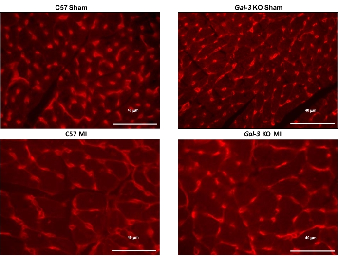
In addition, the hypertrophy of myocytes can be quantified in rhodamine-conjugated lectin-stained slices using the MCSA (Figure 4). The MCSA at 4 weeks showed a significant increase in both genotype groups with MI. However, the increase in hypertrophy observed in C57 mice with MI was absent in the mice lacking Gal-3 (Figure 4). Finally, slices stained with picrosirius red are currently used for quantifying myocardial fibrosis because this stain is specific for collagen fibers and not only for fibrous tissue (unlike Masson's trichome stain). By identifying the collagen fibers (stained in red) and myocytes (stained in yellow), both areas can be quantified, and the collagen percentage can be calculated (Figure 5) either in the infarct zone or the remote zone. The lack of Gal-3 markedly prevented the increase in the collagen volume fraction at the infarct zone in mice with MI (Figure 5).

Figure 1: Macroscopic images of hearts with MI. The left image corresponds to a heart with MI from a C57 mouse, and on the right is a heart from a Gal-3 KO mouse at 7 days post MI. The lack of Gal-3 has disrupted cardiac remodeling. Scale bar = 5 mm. This figure was adapted from Cassaglia et al.4. Abbreviations: MI = myocardial infarction; Gal-3 = galectin-3; KO = knockout. Please click here to view a larger version of this figure.

Figure 2: Echocardiography. (A) Schematic image of the mouse echocardiographic position and the direction of the transductor for the long axis and short axis. Echocardiographic traces were obtained in both strains of mice at 1 week post MI. M-mode echocardiograms of (B) a Gal-3 KO mouse and (C) a control mouse The use of M-mode echocardiography allows for the assessment of the LV wall thickness and the LV internal dimensions in systole and diastole (IVSd, LVIDd, and PWd and IVSs, LVIDs, and PWs are the LV interventricular septum thicknesses and LV posterior wall thicknesses at diastole and systole, respectively.) This figure was adapted from Cassaglia et al.4. Abbreviations: MI = myocardial infarction; Gal-3 = galectin-3; KO = knockout.; LV = left ventricle; IVSd = interventricular septum thickness at diastole; LVIDd = LV internal dimensions at end diastole; PWd = posterior wall thickness at diastole; IVS = interventricular septum; PW = posterior wall; L = long axis; S = short axis. Please click here to view a larger version of this figure.

Figure 3: Representative images of Masson's trichrome-stained slices. The slices are from the middle section of the heart, showing all the measurements obtained for quantifying the infarct size, septum thickness, and scar thickness. These parameters were used to calculate the expansion index (original magnification: 20x). Myocardial morphometry at (A) 1 week after MI in C57 mice; (B) 1 week after MI in Gal-3 KO mice; (C) 4 weeks after MI in C57 mice; (D) and 4 weeks after MI in Gal-3 KO mice. (E,F) TTC staining of infarcted hearts after 24 h in (E) C57 and (F) Gal-3 KO mice. This figure was adapted from Cassaglia et al.4. Scale bars = 1 mm. Abbreviations: MI = myocardial infarction; Gal-3 = galectin-3; KO = knockout. Please click here to view a larger version of this figure.

Figure 4: Representative images of rhodamine-conjugated lectin-stained sections. Rhodamine-stained sections allow for the quantification of the myocyte cross-sectional area at the remote zone at 4 weeks post MI. Original magnification = 400x, scale bar = 40 µm. This figure was adapted from Cassaglia et al.4. Abbreviations: MI = myocardial infarction; Gal-3 = galectin-3; KO = knockout. Please click here to view a larger version of this figure.

Figure 5: Myocardial collagen volume fraction quantification at the remote zone. Data after (A) 1 week and (B) 4 weeks post MI or sham; (C) representative images of a picrosirius red-stained section of the MI zone at 1 week and 4 weeks post MI. The inset shows the representative images obtained with the image analysis software (red shows collagen, and yellow shows non-collagen). Scale bars = 40 µm. This figure was adapted from Cassaglia et al.4. Abbreviations: MI = myocardial infarction; Gal-3 = galectin-3; KO = knockout. Please click here to view a larger version of this figure.
| Experimental groups | C57 | Gal-3 KO | C57 | Gal-3 KO |
| 1 week | 1 week | 4 weeks | 4 weeks | |
| Endocardial Length at MI (mm) | 6.9 | 8.5 | 7.6 | 6.1 |
| Epicardial Length at MI (µm) | 7.1 | 10.7 | 6.7 | 6.5 |
| Endocardial Length non-MI zone (mm) | 5.5 | 3.2 | 6.2 | 4.7 |
| Epicardial Length non-MI zone (mm) | 7.4 | 3.8 | 10.4 | 9.5 |
| Septum Thickness (mm) | 1.1 | 1.1 | 1.4 | 1.1 |
| Scar Thickness (mm) | 0.8 | 0.5 | 0.2 | 0.2 |
Table 1: Representative measurements of the morphometric analysis at 1 week and 4 weeks post MI. Abbreviations: MI = myocardial infarction; Gal-3 = galectin-3; KO = knockout.
Discussion
The experimental model of MI by permanent coronary artery ligature is used for studying a wide variety of pathophysiological mechanisms of cardiac repair and remodeling5,14,17. This article summarizes different methods currently used in this laboratory for studying the temporal evolution of cardiac repair and its effects on post-MI ventricular remodeling14,17. Two models of ischemic cardiopathy have been mostly studied to date: permanent coronary artery ligation, which leads to almost complete necrosis of the area at risk, and transient ischemia followed by reperfusion ("ischemia/reperfusion"), which combines ischemic necrosis with reperfusion injury. Although the latter represents a more translational approach similar to patients receiving reperfusion therapies, this approach has failed to translate results from animal models to patients19.
Another limitation of this method is that, in humans, MI is associated with metabolic dysregulation and arterial atherosclerosis, which differs from rodents, even in models with a high-fat diet. We used the permanent coronary artery ligation model as it is the most appropriate for studying cardiac remodeling after MI. In this protocol, we performed permanent coronary artery ligature in C57 and Gal-3 KO mice, followed by echocardiography at 1 week (not shown) and 4 weeks to evaluate the cardiac remodeling and function in mice. Echocardiography is a non-invasive procedure widely used for studying cardiac remodeling in different cardiovascular diseases. This method allows for the visualization and quantification of cardiovascular structures and the study of cardiac physiology20,21,22.
The temporal evolution of cardiac repair after MI begins with a robust inflammatory response followed by the formation of a fibrous scar and healing, which greatly affects the evolution of whole cardiac remodeling4. Cardiac remodeling after MI is characterized by thinning of the MI area followed by fibrosis and hypertrophy of the non-infarcted myocardium, ventricular dilation, and heart failure. Therefore, the recognition of the anatomical and histological changes during the evolution of the infarction allows for better characterizing the remodeling. Thus, slices stained with Masson's trichrome can be used for measuring the infarct size and the early remodeling, characterized by the expansion of the infarct zone. In the days to weeks that follow an acute MI, the infarcted area suffers a radial thinning and a circumferential increase, which is referred to as the expansion of the MI zone.
Here, we have shown how to quantify the expansion in the infarct zone in slices stained with Masson's trichrome. In addition, in slices stained with picrosirius red, we have shown how to evaluate the amount of collagen in the infarct and remote zones5,17. The replacement of necrotic tissue by fibrosis is clearly a consequence of the cardiac repair, and any change in the amount of collagen reflects an alteration in the healing process of the infarct and affects the early remodeling of the scar. The same slices allow the definition of the extracellular matrix remodeling of the non-infarct zone4,17. Finally, in rhodamine-stained sections, the quantification of the MCSA is useful to evaluate the remodeling of the infarct zone, which may be counterproductive and increase myocardial stiffness, leading to heart failure. A collagen-rich scar is necessary to prevent the rupture of the infarct area, while interstitial fibrosis is detrimental. In conclusion, the temporal evolution of myocardial infarction is a useful model for studying the effects of cardiac repair on the temporal evolution of cardiac remodeling. Thus, the methods described above would allow for the comparison of two genotypes for evaluating the consequences of inflammatory interventions on cardiac repair and cardiac remodeling in mice.
Disclosures
The authors have no conflicts of interest to declare.
Acknowledgements
The authors gratefully appreciate the technical assistance of Ana Chiaro. This work was supported by grants from the Argentinean Agency for Promotion of Science and Technology (PICT 2014-2320, 2019-02987 and PICT 2018-03267 to VM) and the University of Buenos Aires (UBACyT 2018- 382 20020170100619BA to GEG).
Materials
| Name | Company | Catalog Number | Comments |
| 8-0 silk suture | Ethicon | ||
| C57BL/6J mice | Department of Bioresources of the Faculty of Veterinary of the University of Buenos Aires, Argentina | ||
| Forceps | |||
| Hardvard 386 respirator | Hardvard company | ||
| Heating pad | maintain animal's temperature during surgery | ||
| Image Pro-Plus 6.0 | Media Cybernetics | Image Analysis Software | |
| Ketamine | Holiday | ||
| Masson Trichrome | BIOPUR | ||
| Picrosirius red | BIOPUR | ||
| Retractors | |||
| Rodent Ventilator Model 683 | Harvard Apparatus | Mechanical ventilator | |
| Scissors | |||
| Stereoscopic magnifying glass | Arcano | ||
| Vivid 7 machine (General Electric Medical Systems, Horten, Norway) | General Electric | Any tracking software can be utilized with this protocol | |
| WGA no. RL-1022, Vector Laboratories, Burlingame | Vector Laboratories | ||
| Xylazine | Pro-Ser |
References
- Opie, L. H., Commerford, P. J., Gersh, B. J., Pfeffer, M. A. Controversies in ventricular remodelling. Lancet. 367 (9507), 356-367 (2006).
- Frangogiannis, N. G. The inflammatory response in myocardial injury, repair, and remodelling. Nature Reviews Cardiology. 11 (5), 255-265 (2014).
- Clarke, S. A., Richardson, W. J., Holmes, J. W. Modifying the mechanics of healing infarcts: Is better the enemy of good. Journal of Molecular and Cellular Cardiology. 93, 115-124 (2016).
- Cassaglia, P., et al. Genetic deletion of galectin-3 alters the temporal evolution of macrophage infiltration and healing affecting the cardiac remodeling and function after myocardial infarction in mice. American Journal of Pathology. 190 (9), 1789-1800 (2020).
- Seropian, I. M., et al. Galectin-1 controls cardiac inflammation and ventricular remodeling during acute myocardial infarction. American Journal of Pathology. 182 (1), 29-40 (2013).
- Yang, R. Y., Rabinovich, G. A., Liu, F. T. Galectins: Structure, function and therapeutic potential. Expert Reviews in Molecular Medicine. 10, (2008).
- Liu, Y. H., et al. N-acetyl-seryl-aspartyl-lysyl-proline prevents cardiac remodeling and dysfunction induced by galectin-3, a mammalian adhesion/growth-regulatory lectin. American Journal of Physiology-Heart and Circulatory Physiology. 296 (2), H404-H412 (2009).
- Ibarrola, J., et al. Myocardial injury after ischemia/reperfusion is attenuated by pharmacological galectin-3 inhibition. Scientific Reports. 9, 9607 (2019).
- de Boer, R. A., et al. Predictive value of plasma galectin-3 levels in heart failure with reduced and preserved ejection fraction. Annals of Medicine. 43 (1), 60-68 (2011).
- Li, S., Li, S., Hao, X., Zhang, Y., Deng, W. Perindopril and a galectin-3 inhibitor improve ischemic heart failure in rabbits by reducing Gal-3 expression and myocardial fibrosis. Frontiers in Physiology. 10, 267 (2019).
- Mo, D., et al. Cardioprotective effects of galectin-3 inhibition against ischemia/reperfusion injury. European Journal of Pharmacology. 863, 172701 (2019).
- Suthahar, N., et al. Galectin-3 activation and inhibition in heart failure and cardiovascular disease: An update. Theranostics. 8 (3), 593-609 (2018).
- US Committee for the Update of the Guide for the Care and Use of Laboratory Animals. Guide for the Care and Use of Laboratory Animals. National Academies Press. , (2011).
- González, G. E., et al. Galectin-3 is essential for early wound healing and ventricular remodeling after myocardial infarction in mice. International Journal of Cardiology. 176 (3), 1423-1425 (2014).
- González, G. E., et al. Cardiac-deleterious role of galectin-3 in chronic angiotensin II-induced hypertension. American Journal of Physiology-Heart and Circulatory Physiology. 311 (5), H1287-H1296 (2016).
- González, G. E., et al. Effect of early versus late AT-1 receptor blockade with losartan on postmyocardial infarction ventricular remodeling in rabbits. American Journal of Physiology-Heart and Circulatory Physiology. 297 (1), H375-H386 (2009).
- Muthuramu, I., Lox, M., Jacobs, F., De Geest, B. Permanent ligation of the left anterior descending coronary artery in mice: A model of post-myocardial infarction remodelling and heart failure. Journal of Visualized Experiments. 94 (94), (2014).
- Dai, W., Wold, L. E., Dow, J. S., Kloner, R. A. Thickening of the infarcted wall by collagen injection improves left ventricular function in rats: A novel approach to preserve cardiac function after myocardial infarction. Journal of the American College of Cardiology. 46 (4), 714-719 (2005).
- Jones, S. P., et al. The NHLBI-sponsored Consortium for preclinicAl assESsment of cARdioprotective therapies (CAESAR): A new paradigm for rigorous, accurate, and reproducible evaluation of putative infarct-sparing interventions in mice, rabbits, and pigs. Circulation Research. 116 (4), 572-586 (2015).
- Gao, S., Ho, D., Vatner, D. E., Vatner, S. F. Echocardiography in mice. Current Protocols in Mouse Biology. 1, 71-83 (2011).
- Yang, X. P., et al. Echocardiographic assessment of cardiac function in conscious and anesthetized mice. American Journal of Physiology. 277 (5), H1967-H1974 (1999).
- Lindsey, M. L., Kassiri, Z., Virag, J. A. I., Castro Brás, d. e., E, L., Scherrer-Crosbie, M. Guidelines for measuring cardiac physiology in mice. American Journal of Physiology-Heart and Circulatory Physiology. 314 (4), H733-H752 (2018).
Reprints and Permissions
Request permission to reuse the text or figures of this JoVE article
Request PermissionThis article has been published
Video Coming Soon
Copyright © 2025 MyJoVE Corporation. All rights reserved